Figure 3.
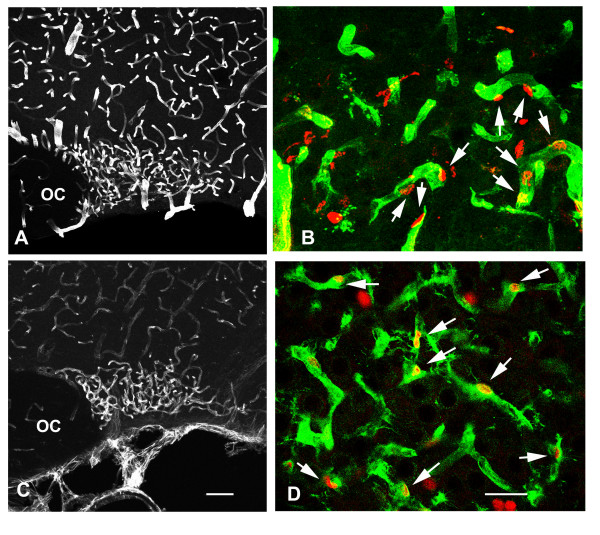
Cell proliferation mostly concerns SON capillary endothelial cells. BrdU was administrated to osmotically stimulated rats drinking 2% saline during 6 days, and the animals were fixed 5 hours after the BrdU administration. A, C: Stack confocal images (10 μm-thick) of sections labeled for endothelial cell markers. EBA immunostaining is associated with vessels located within the SON and the surrounding regions (A), whereas nestin immunostaining is associated with SON vessels, but only faintly labels those vessels located in the surrounding regions (C). B, D: Merged confocal images of sections double labeled for BrdU and for one of the two the endothelial cell markers. Throughout the SON, BrdU-labeled nuclei are frequently associated with vessel structures labeled for EBA (arrows in B) or nestin (arrows in D). EBA: endothelial brain antigen; OC: optic chiasma. Scale bars: A, C = 100 μm; B, D = 25 μm.

