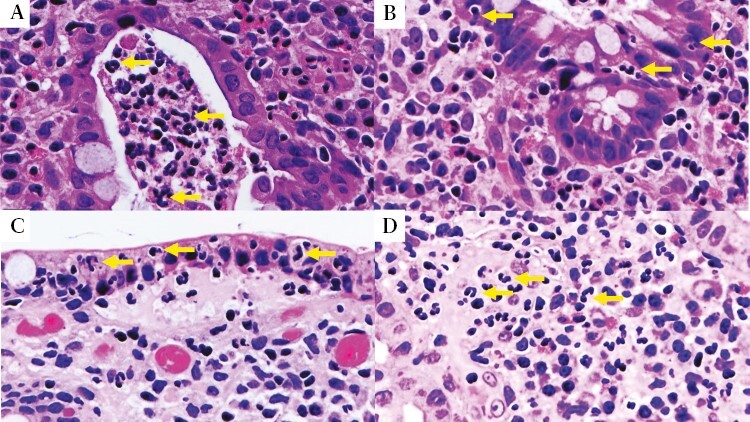
Figure 1.

Examples of neutrophilic infiltration. A] Crypt abscess [arrows on neutrophils] H&E × 40. B] Neutrophils infiltrating the cryptal epithelium [arrows on neutrophils] H&E × 40. C] Neutrophils in the superficial epithelium [arrows on neutrophils] H&E × 40. D] Neutrophils in lamina propria [arrows on neutrophils] H&E × 40. H&E, haematoxylin and eosin.
