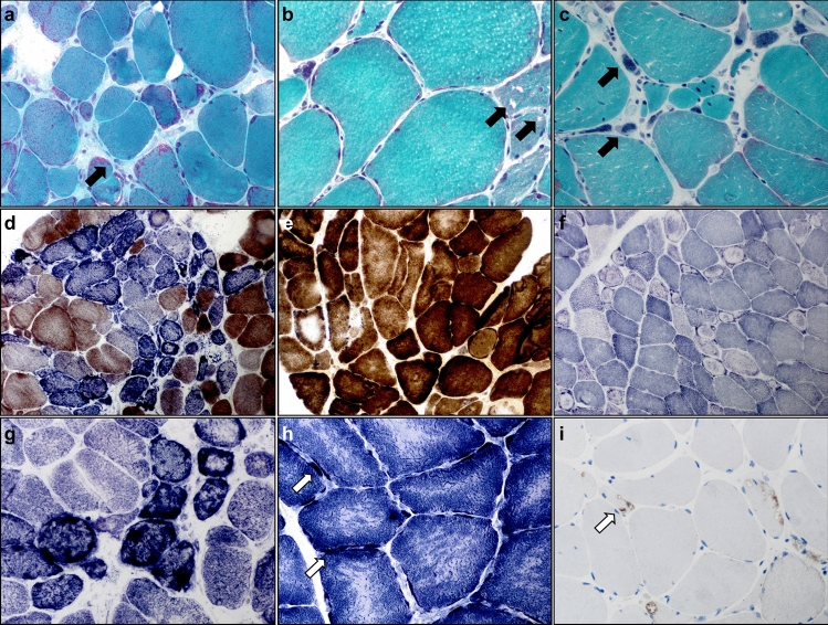
Fig. 2.
Histomorphological alterations of skeletal muscles from DM2 patients. Photomicrographs a–i illustrate histological findings in individual patients with DM2. a–c (Gömöri-trichrome staining): ragged red and split fibers (black arrow) (a; × 400), rimmed (black arrow) and non-rimmed vacuoles (b; × 400), grape-like nuclear clumps/bags (black arrow) (c; × 400). d, e (COX–SDH enzyme histochemical preparation): abundant and focally clustered COX-negative SDH-positive fibers (d; × 200), central displacement and variable irregularity of COX-activity in some fibers (e; × 400). f (NADH-TR staining): many myofibers showing ‘ringbinden’ and ring fibers mostly in type 2 fibers, while dark type 1 fibers only rarely show them (× 200). g–h (SDH staining): focally clustered ragged blue fibers (g, × 400), targetoid-like lesions in fibers with subsarcolemmal substrate accumulation (arrows) (h; × 600). i (p62 immunohistochemistry): autophagolysosomal aggregates (arrow) (× 400)

