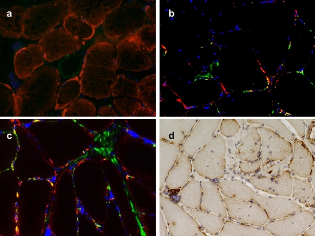
Fig. 4.

Auto- and mitophagy in DM2 muscle biopsy samples. a–c Representative immunofluorescence of individual biopsy samples derived from DM2 patients with co-localization of mitochondrial marker protein TOM20 (a; red) with LC-3 (green); TOM20 (b; red) with p62 (green); and COX-IV (c; red) with p62 (green). DAPI staining of nuclei in blue (b, c; magnification × 400). While subsarcolemmal accumulated mitochondria are visible by immunofluorescence, co-staining (yellow) is only evident for p62 and TOM20/COX-IV (b, c). d Immunohistochemistry for the mitophagy marker BNIP3 shows a strong staining reaction in the subsarcolemmal region in many myofibers (× 200)
