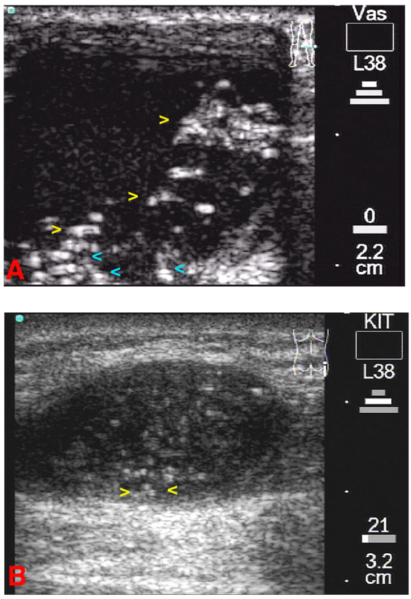
Figure 4.
4A: Transverse scan of the patient's left knee. A large cystic onchocercoma can be seen. Yellow arrowheads indicate at moving fragments of the worm(s), while the blue arrowheads indicates static fragments of the worm body (ies). The corresponding video image can be seen as Additional File 3. 4B: Longitudinal scan of the patient's right trochanter. A medium sized cystic nodule where minimal movements were detected. The worm(s) is (are) surrounded by cystic fluid seen as echo-free areas (yellow arrowheads) and can be differentiated from the capsule of the onchocercoma.

