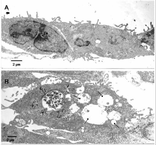
Figure 8.

Transmission electron microscope of A549 cells. A = control cells present numerous microvilli and the normal cytoplasmic inclusions. B = a vacuolised cytoplasm (arrows) after exposition to TD organic extract. This modified morphology of the cytoplasm is frequent in cells treated with 75 μg/ml TD organic extract at 72 h. (bar = 2 μm).
