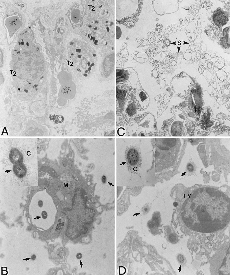
FIG. 2.
Electron microscopy of lungs of mice infected with 107 S. pneumoniae cells and sacrificed 72 h later. Type II pneumocytes (T2) proliferated after infection (A; magnification, ×9,600) and secreted abnormal amounts of surfactant (S) in alveoli (C; ×6,000). Although S. pneumoniae cells (arrows) were partly eradicated through phagocytosis (M, macrophage) (B; ×18,000), extracellular killing also seemed to occur, as the polysaccharide capsule (C) of bacteria localized outside phagocytes in areas of intense inflammation appeared more disaggregated (thinner and more diffuse) (B [arrows] and inset; ×44,000) than the capsule of bacteria localized in less severely inflamed areas (D [×18,000] and inset [×44,000]). LY, lymphocyte.

