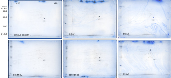
Figure 4.
2D VOPBA of sodium deoxycholate soluble proteins after β octyl-glucopyranoside extraction of PS Clone D cell monolayer. The antigen preparation used in the VOPBA staining of the 2D blots is labelled on the bottom left of each blot. Two images are superimposed in each panel. The Ponceau S scan of each blot is shown in greyscale and shows the universe of spots transferred to the blot. The spots reactive in the VOPBA analysis are shown in blue. Spots marked with a circle were identified as LAMR1 and the spot in the DENV4 blot marked with a rectangle was identified as lamin B1.

