



Abstract
Aging results in remodeling of T cell immunity and is associated with poor clinical outcome in age-related diseases such as cancer. Amongst the hallmarks of aging, changes in host and cellular metabolism critically affect the development, maintenance, and function of T cells. Although metabolic perturbations impact anti-tumor T cell responses, the link between age-associated metabolic dysfunction and anti-tumor immunity remains unclear. In this review, we summarize recent advances in our understanding of aged T cell metabolism, with a focus on the bioenergetic and immunologic features of T cell subsets unique to the aging process. We also survey insights into mechanisms of metabolic T cell dysfunction in aging and discuss the impacts of aging on the efficacy of cancer immunotherapy. As the average life expectancy continues to increase, understanding the interplay between age-related metabolic reprogramming and maladaptive T cell immunity, will be instrumental for development of therapeutic strategies for older patients.
I. Introduction
The demographics of the human population is aging in an unprecedented manner. It is estimated that by the middle of the 21st century, nearly one-quarter of the world population will consist of adults 65 years of age or older.1 In addition to being associated with the development of multiple comorbidities, including diabetes, autoimmune disease, neurological deficits, and an increased susceptibility to infectious agents, aging is the most well-known risk factor for the development of cancer, the incidence of which disproportionately increases with age. It is now appreciated that functional dysregulation of the immune system is a central feature of the aging process. Surprisingly, our knowledge of the aging immune system, the mechanistic drivers of immune dysfunction during aging and the impact of the aged immune system on age-associated diseases remains incomplete.
The vertebrate immune system is equipped with a diverse cellular arsenal, capable of providing highly specialized and durable protection against various pathogens. Innate immunity provides immediate protection against invading pathogens and relies on a limited but evolutionary conserved set of germline-encoded receptors that recognize common constituents of microbes. Despite being rapid in nature, innate immune responses are limited in specificity, diversity and memory. By contrast, adaptive immune responses are highly specialized and rely on a diverse pool of lymphocytes capable of recognizing and responding to a large number of microbial and other antigens. The generation of a diverse adaptive immune repertoire is enabled by the rearrangement of antigen receptor genes resulting in different T and B lymphocyte clones with unique receptor specificities.
T lymphocytes originate from hematopoietic stem cell (HSC)-derived lymphoid progenitor cells in the bone marrow (Figure 1). These lymphoid progenitors migrate to the thymus, a specialized primary lymphoid organ critically involved in T cell development, and further mature into cytotoxic or helper T cell subsets. Upon thymic egress, mature T cells migrate into secondary lymphoid organs, where they continuously survey peptide fragments presented by major histocompatibility complex (MHC) molecules on the surface of antigen-presenting cells (APCs). Naïve T cell activation in the lymph node requires two signals: Ligation of the T cell receptor (TCR) by cognate peptide-MHC initiates naive T cell activation (signal one), and results in a downstream signaling cascade that promotes T cell proliferation, differentiation, and effector functions. Simultaneous engagement of the costimulatory receptor CD28 by its ligands B7–1 or -2 (CD80 or CD86) (signal two) is required for full T cell activation initiated by the TCR. In addition to these two signals, signal three provided by inflammatory cytokines including IL-12 and type I IFN promotes T cell differentiation and acquisition of effector functions. These signals culminate in the differentiation of activated T cells into specialized killer or helper T cell subsets (Figure 1).2–4 While CD8+ T cells produce effector molecules including perforins and granzymes to directly induce destruction of target cells, subsets of CD4+ T cells play an important role in fine-tuning immune responses through the secretion of specialized cytokines and by engaging in interactions with various immune cells.
Figure 1: A brief overview of T cell development and activation.

T cell development begins in the bone marrow with hematopoietic stem cells that acquire a lymphoid progenitor state. These cells migrate to the thymus, a specialized primary lymphoid organ, to undergo additional maturation and selection against self-reactive T cell clones. During this process, progenitor T cells differentiate into either mature CD4+ or CD8+ T cells and exit the thymus. These antigen-naive T cells can be activated in the lymph node by dendritic cells which provide TCR, co-stimulatory and cytokine signaling for the acquisition of effector T cell functions.
Although much of our understanding of T cell immunity originated from studies utilizing immunocompetent young hosts, studies in aged model systems have taught us that T cell function and phenotype are considerably altered with age.5 The complex biological changes associated with aging impact multiple facets of T cell immunobiology including development, activation, differentiation, and survival. Age-associated deficits in T cell responses against cancer, infectious and autoimmune diseases are well documented in elderly demographics. Recent studies have revealed that T cells a well as B cells play an important role in shaping the immune response to severe acute respiratory syndrome coronavirus 2 (SARS-CoV-2) infections.6 Furthermore, one of the most critical determinants of disease severity was age,7 and studies have highlighted the crucial role of age-associated T cell dysregulation in the perpetuation and aggravation of disease severity.8 However, the mechanisms driving age-associated T cell dysfunction and disease progression remain an active area of investigation.
Impaired T cell function in aged hosts is attributed to the cumulative deficits in cellular and physiological integrity caused by molecular hallmarks of aging. Genomic instability, telomere attrition, epigenetic alterations, loss of proteostasis, cellular senescence and numerous other biological deficits all contribute to cellular and physiological decline.9 Amongst these perturbations, one of the most prominent hallmarks of aging involves metabolic dysfunction in which chronic dysregulation of cellular and host bioenergetic programs results in altered cellular function and increased susceptibility to metabolic disorders, including obesity, type II diabetes and non-alcoholic fatty liver disease.10 These metabolic adaptations may be driven by age-associated defects in mitochondria, which are multi-faceted organelles that play a critical and non-redundant role in the production of ATP, maintenance of calcium homeostasis and regulation of cellular apoptosis.11 Age-associated mitochondrial dysfunction results in impaired mitochondrial respiration and mitophagy, augmentation of reactive oxygen species (ROS) production, and increased apoptosis in inflammatory settings. The relationships between mitochondrial degeneration and aging have been the subject of many recent investigations.12
Age-associated mitochondrial and metabolic reprogramming has a direct impact on the immune system and its role in diseases such as cancer, autoimmunity and chronic infections. In particular, the field of T cell metabolism has become an area of substantial investigation over the past decade. Recent work highlights the importance of cellular bioenergetic programs in peripheral T cell maintenance, activation, differentiation, and exhaustion. However, the metabolic pathways which control the fate of T cells are often rewired as a result of age-associated mitochondrial and metabolic perturbations.13 In this review, we summarize past progress and recent advances in our understanding of the role of aging in T cell immunity and metabolism, with emphasis on the implications of metabolic reprogramming of aged T cells in regulating anti-tumor immunity.
II. Early studies of T cell aging
The notion that aging impairs T cell development and function is a long-standing observation. Studies in the 1960’s revealed that aging negatively impacts the activation and distribution of T cells. In humans, both the frequency and absolute quantity of spontaneous sheep red blood-cell (SRBC) rosette-forming T lymphocytes decreased with age,14 and was correlated with an overall decline in thymosin, a 5-Da polypeptide hormone critically involved in T cell development and function.15 These findings were consistent with impairment of delayed hypersensitivity responses (DHR) in aged individuals upon antigenic sensitization with 2,4-dinitrochlorobenzene (DNCB).16,17 Furthermore, in vitro stimulation of lymphocytes using mitogens such as phytohemagglutinin (PHA) resulted in attenuated T cell proliferative responses in aged individuals.18–20 Similar numerical and functional T cell deficits have been documented in aged mice.21–24 Studies have highlighted the potential implications of age-associated T cell dysfunction in various disease settings,25–27 underscoring a need to understand the cellular and molecular mechanisms involved.
Building upon the previously acknowledged impact of aging on T cell immunity, recent studies have integrated more sophisticated tools such as single-cell DNA/RNA sequencing to further profile the systemic effects of aging on the immune system.28,29 These efforts, along with many other studies, have provided greater insight into how aging modulates T cells. It is now appreciated that numerous age-associated biological alterations may contribute to a defective T cell response.5 However, the biological mechanisms underlying age-associated T cell dysfunction remain unclear and context dependent.
III. T cell development
One of the most striking features of aging is thymic involution, during which the thymus degenerates over time and no longer replenishes naïve CD8+ and CD4+ T cell pools necessary for protective immunity.30–32 Impaired naïve T cell development disrupts the composition of peripheral T cells, resulting in compensatory expansion of pre-existing T cell clones. These adaptations lead to decreased TCR diversity, an altered distribution of T cell subsets, and qualitative changes in T cell responses following TCR stimulation. These cellular changes and clonal expansion may contribute to the loss of immune responses to a diverse set of antigens in the aged.
In early studies, thymectomy of young C57BL/6 and A/J mice accelerated the decline in cellular immunity and precipitated the generation of dysfunctional T cell populations characterized by attenuated responsiveness to PHA and staphylococcal enterotoxin B (SEB), similar to those observed in aged mice.24 Consistent with this finding, altered lymphocyte sub-type distribution and impaired responsiveness to mitogenic signaling is observed in patients following thymectomy.24,33 Species-specific adaptation to thymic involution also has been reported. In contrast to mice, which predominantly rely on thymically-derived T cells to replenish the peripheral naïve T cell pool, peripheral expansion of naïve T cells in aged humans was sufficient to compensate for thymic involution.34–36 This divergence suggests that mouse studies may offer limited insight into the processes governing adaptation to thymic involution in humans and the overall impact of such adaptation on the human aging process. While the use of aged mice as an experimental model system will continue to guide our basic understanding of immune aging, the integration of human studies, including use of primary human cells and grafts, will be necessary for the validation of mechanistic insights obtained from animal models.
Although several mechanisms have been implicated in age-associated thymic involution, the central driver of thymic attrition remains unclear. The process of aging modifies hematopoietic reprogramming of precursor cells in the bone marrow to favor commitment towards myeloid over lymphoid lineages37 which may contribute to reduced thymic output. However, thymic atrophy is initiated prior to alterations in the hematopoietic compartment. Studies have demonstrated that age-associated thymic atrophy in healthy hosts is associated with alterations in thymic stromal cells38,39 and that attenuated H2O2-reducing enzyme and mitochondrial dysfunction of stromal cells may contribute to thymic atrophy.40 Consistent with this finding, genetic and biochemical augmentation of anti-oxidants was shown to ameliorate thymic atrophy.40
The metabolic environment plays a major role in thymic atrophy and generation of naïve T cells. For example, age-associated thymic involution correlates with accretion of lipid-laden adipocytes, while disrupting accumulation of thymic adipocytes through caloric restriction attenuates tissue atrophy.41 A recent study by Spadaro et al. demonstrated that caloric restriction in humans is associated with impaired expression of platelet activating factor acetyl hydrolase (PLA2G7), increased mitochondrial biogenesis and PPAR-α-driven lipid oxidation in thymic adipocytes. Deletion of PLA2G7 in mice resulted in decreased thymic lipoatrophy.42 Consistent with the deleterious effects of age-associated adipocyte remodeling of thymic tissue architecture, high-fat diet (HFD)-induced obesity accelerated thymic involution, reduced peripheral naïve T cell numbers and decreased TCR repertoire diversity in mice,43 while progressive adiposity in mid-aged humans reduced thymic output.43 Similarly, severe thymic atrophy was observed in leptin-deficient obese mice in which reconstitution of leptin reversed obesity and thymic involution.44,45 In contrast to these findings, other work suggests that thymic size, cell number and output may increase in HFD-induced obese mice,46 as a result of the role of leptin in augmenting thymocytes and T cell function.47,48 Additional studies are needed to better understand mechanisms connecting nutrient signaling with T cell development during aging. In summary, age-associated metabolic alterations significantly impact thymic structural integrity and T cell development, which further impact T cell responses required for protective immunity.
IV. Naïve T cell homeostasis
In the periphery of aged hosts, pre-existing T cells undergo additional expansion to compensate for impaired naïve T cell development. During this process, aging exerts distinct effects across pre-existing naïve CD8+ and CD4+ T cells. For reasons incompletely understood, naïve CD8+ T cells exhibit greater vulnerabilities to the effects of aging in comparison to CD4+ T cells.49–53 Consistent with this finding, one of the cardinal features of aging in healthy adults is reduced naïve CD8+ T cell numbers in the periphery.54 The presence of age-associated “memory T cells with a naïve phenotype” (TMNP cells) in the naïve CD8+ T cell pool suggests that the number of naïve CD8+ T cells is likely overestimated in most studies which do not utilize TMNP markers such as CD49b and CXCR3 for additional segregation.55 Overall, the aging process preferentially decreases the number and longevity of CD8+ T cells despite increasing the longevity of naïve CD4+ T cells.56 These differences may be explained by the presence of naïve-like CD45RA+CD25dimCD4+ T cells in aged mice, which are characterized by increased sensitivity to IL-2.51 Although the consequences of aging on naïve CD4+ T cell function remain unclear in vivo, several studies have demonstrated defective proliferation and impaired CD25 expression in aged naïve CD4+ T cells following TCR engagement in vitro.56,57
Defects in the naïve CD8+ T cell pool can be attributed to cell-extrinsic and -intrinsic changes that accompany the aging process. Cell-intrinsic factors, including genetic, epigenetic, and metabolic alterations, cumulatively contribute to the overall dysfunction of naïve CD8+ T cells. Telomere length is significantly reduced in the naïve CD8+ T cell compartment, likely as consequence of heightened homeostatic proliferation in aged hosts.58,59 Telomere shortening of CD8+ T cells is augmented with age-associated elevation of mitochondrial stress and ROS accumulation. Mitochondrial dysfunction-induced telomere damage was reversed with the use of a ROS scavenger.60 Decreased expression of the double-strand break repaired nuclease MRE11A causes telomeric damage and accelerates the aging process in T cells from patients with rheumatoid arthritis.61 Telomere shortening disrupts T cell homeostasis and survival, as illustrated by the finding that telomerase mutation carriers with short telomere length develop T cell immunodeficiency.62 Conversely, increased telomere length and telomerase activity in T cells has been associated with health benefits in healthy centenarians.63
Age-associated epigenetic modifications also contribute to remodeling of the naïve CD8+ T cell compartment. Aged naïve CD8+ T cells exhibit differential chromatin accessibility profiles compared to aged naïve CD4+ T cells.52,64 Transcriptional activity of NFκB, STAT, YY1, TCF1 and NRF1, all of which play key functions in T cell proliferation, were reduced in aged naïve CD8+ T cells.52,64,65 For example, aged naïve CD8+ T cells exhibit decreased binding of NRF1 to its motif as a result of limited chromatin accessibility.66 With the transcriptional coactivator PGC-1α, NRF1 promotes transcription of genes involved in glutathione biosynthesis,67,68 the electron transport chain (ETC),69 and positively regulate the mitochondrial transcription factor A (TFAM),70 all of which play critical roles in mitochondrial biogenesis and respiration. Consistent with these data, basal and maximal oxygen consumption rates (OCR) were decreased in aged naïve CD8+ T cells despite observations of increased mitochondrial mass.66 Naïve CD8+ T cells heavily rely on oxidative phosphorylation (OXPHOS) for homeostatic expansion, and age-associated epigenetic regulation of NRF1 and genes associated with mitochondrial respiration likely contribute to defective homeostatic proliferation of aged naïve CD8+ T cells. Lastly, although epigenetic changes directly impact T cell metabolism, metabolic changes also influence the epigenetic states of T cells, ultimately shaping T cell fate decisions and differentiation programs.71–74 However, it remains unclear whether metabolic changes contribute to altered epigenetic states in aged T cells.
Post-transcriptional regulators, including microRNAs, also play an important role in aged-associated naïve T cell remodeling. In particular, age-dependent reconfiguration of microRNA networks predicted a major defect in FoxO1 signaling,75 which has been reported to repress T cell senescence and promote proteostasis.76,77 Furthermore, FoxO1 directly promotes expression of the interleukin 7 (IL-7) receptor,75 which plays an important role in maintaining naïve CD8+ T cell homeostasis.78 Consistent with this finding, IL-7R expression was reduced in naïve CD8+ T cells from aged hosts and inhibition of FoxO1 in young naïve CD8+ T cells decreased IL-7R expression to levels observed in aged T cells.75 Although IL-7 modulates glucose uptake and glycolytic flux, reports on the role of IL-7 in the regulation of Glut1 expression are conflicting.79,80 IL-7 also increases the expression of CD98, a heterodimer of SLC3A2 and SLC7A5 which forms the large neutral amino acid transporter, to sustain IL-7 induced T cell growth.81 While age-associated changes in metabolic, genetic, epigenetic, and post-transcriptional regulation have potential to contribute to defective expansion of naïve CD8+ T cells in aged hosts, there is still a need to investigate how these changes impact the function of aged T cells to alter immune control in vivo.
V. T cell activation & metabolism
Naïve T cell activation is a highly regulated process which relies on appropriate integration of TCR, co-stimulatory and cytokine signaling. Early studies suggested that naïve T cell activation becomes dysfunctional with age, as evidenced by decreased proliferation in response to T cell stimulation.18–20 Although the consequences of aging on early events following TCR stimulation are poorly characterized for both CD8+ and CD4+ T cells, several distinct molecular and cellular features of aged T cell activation have been described. Activation of aged naïve T cells is associated with impaired immune synapse formation,82 recruitment of phosphorylated Lck and LAT to the lipid raft,83 Ca2+ flux,84,85 cytokine production, and expression of activation markers such as CD25 and CD69.86 Moreover, TCR engagement of aged T cells is associated with increased apoptosis and DNA damage response.87,88 As previously described, prior to TCR engagement, aged naïve CD8+ T cells display baseline alterations in genetic, epigenetic, and post-transcriptional regulatory networks which have important implications for T cell activation. For example, the microRNA mir-181a controls TCR activation threshold during thymic selection and peripheral T cell activation. Loss of miR-181a expression during aging results from the age-associated decline of YY1, a transcription factor that promotes miR-181a expression by controlling pri-miR-181a/b1 enhancer activity.89 In addition to baseline alterations in TCR signaling, metabolic perturbations also contribute to defective cellular activation.
Metabolic requirements for T cell activation
Metabolic reprogramming following T cell activation is indispensable for maintenance of high energetic demands resulting from rapid proliferation, differentiation and acquisition of effector T cell function.90 Naïve T cells exist in a state of quiescence characterized by low metabolic activity and preferential use of OXPHOS for the generation of ATP. Following TCR engagement, naïve T cells rapidly switch towards the use of aerobic glycolysis, with a reliance on glutaminolysis, pentose-phosphate pathway activity, and 1-carbon metabolism to increase energy production.91–93 While numerous bioenergetic pathways are actively utilized during T cell activation, we focus here mainly on glycolytic and mitochondrial metabolism which are highly altered with aging (Figure 2).
Figure 2: Naïve T cells from aged hosts display attenuated glycolysis and mitochondrial metabolism.
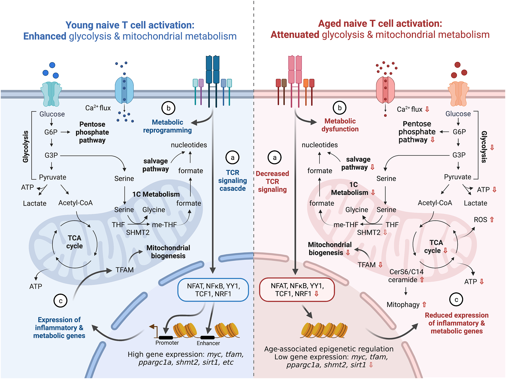
Aged naïve T cells display defective TCR signaling (a), early metabolic activities (including glycolysis and pentose phosphate pathway) (b), and transcriptional and proteomic reprogramming required for mitochondrial biogenesis (c).
Early TCR stimulation events induce aerobic glycolysis, leading to rapid production of ATP and cytokine production.94,95 Aerobic glycolysis involves the activation of pyruvate dehydrogenase kinase 1 (PDHK1) which phosphorylates and deactivates pyruvate dehydrogenase (PDH). Increased PDHK1 and subsequently decreased PDH activity in T cells lead to attenuated conversion of pyruvate into acetyl-coenzyme A for mitochondrial import and increased conversion of pyruvate into lactate, catalyzed by lactate dehydrogenase (LDH).95 This metabolic switch disengages the binding of LDH to the 3’ untranslated region (UTR) of IFN-γ, IL-2 and TNF-α mRNA and thereby increases cytokine production. Lactate dehydrogenase A (LDHA) also controls IFN-γ expression through an alternative mechanism involving maintenance of acetyl-CoA, which enhances histone acetylation and transcription of IFN-γ mRNA.96 Glyceraldehyde 3-phosphate dehydrogenase (GAPDH) is another crucial enzyme upstream of the glycolytic pathway which binds to the 3’ UTR of IFN-γ mRNA prior to T cell activation, and TCR-mediated glycolysis disengages GAPDH-IFN-γ mRNA binding to promote IFN-γ translation.93 In conjunction with regulating cytokine production, glycolysis plays a crucial role in the generation of metabolic fuels required for optimal T cell activation, including the biosynthesis of nucleotides, amino acids, proteins and fatty acids.97 Glycolysis and glutaminolysis were also shown to co-operatively deprive the hexosamine pathway of glucose and glutamine, which inhibits N-glycan branching and favors the growth and differentiation of pro-inflammatory Th17 cells.98,99 Lastly, glycolysis increases levels of acetyl-CoA and promotes histone acetylation, thereby enabling the generation of open and permissive chromatic structures required for T cell activation.73,100 Together, the glycolytic pathway initiates metabolic reprogramming to enable rapid cytokine production, proliferation, and activation of T cells.97,101,102
Upon TCR-stimulation, mitochondria buffer Ca2+ flux in the cytosol.103,104 Mitochondrial localization proximal to the immune synapse decreases local cytosolic Ca2+ and calcium-dependent channel inactivation, thus augmenting overall calcium influx.105–107 Uptake of Ca2+ into the mitochondrial matrix enhances the activity of proteins associated with TCA cycle, and promotes OXPHOS, ATP generation and ROS production, all of which have an important role during T cell activation.106,108 The initial Ca2+ flux followed by Ca2+ influx from the ER and extracellular compartment activate calcineurin, which leads to the activation of NFAT. Mitochondrial production of ROS plays an important role in inducing NFAT-dependent Myc expression, which augments T cell proliferation and metabolic reprogramming.91,108 In particular, Myc-dependent metabolic reprogramming involves augmentation of glycolysis and glutaminolysis.91
Mitochondrial biogenesis and remodeling accompany TCR stimulation, and is accompanied by remodeling of the mitochondrial proteome and augmentation of 1-carbon metabolism.92,109 Mitochondrial respiration and ATP production also play a critical role in supporting energy demands following T cell activation and inhibition of ATP synthase using oligomycin blunts T cell activation.93
During mitochondrial biogenesis, mitochondria are rewired to become anabolic engines. One-carbon metabolism utilizes serine to generate one-carbon units for thymidine and purine biosynthesis, which play an important role in early T cell activation and proliferation (Figure 2).92,110 During this process, serine is converted to glycine in the cytosol and mitochondrial matrix by serine hydroxymethyltransferase-1 and 2 (SHMT1 and 2), respectively. In addition, serine-driven one-carbon metabolism contributes to the maintenance of redox homeostasis through the generation of glycine for glutathione synthesis and NADPH, which supports antioxidant defence and reductive biosynthesis.92,111 In summary, a metabolic switch towards aerobic glycolysis provides T cells with initial energy and cues required for rapid cellular proliferation, but mitochondrial metabolism is required for sustaining T cell activation.
Age-associated metabolic changes in T cell activation
Although the literature broadly suggests that age-associated metabolic dysfunction may contribute to an altered T cell response, it remains unclear precisely how age-associated metabolic alterations impact the bioenergetic program of T cells at discrete phases of development and activation. Here, we focus on age-associated metabolic alterations upon naïve T cell activation.
Alterations in glycolysis correlate with age-associated T cell dysfunction. Early studies suggested that LDH activity was significantly reduced in aged T cells and correlated with decreased T cell proliferation.33,112 Consistent with these studies, activation of naïve aged CD4+ T cells resulted in attenuated ECAR and reduction in metabolites associated with glycolysis and the pentose phosphate pathway (Figure 2).57 It remains unclear whether glycolysis is also impaired in naïve CD8+ T cells during aging. Reduced glycolysis in CD4+ T cells may result from age-associated changes in the TCR signaling threshold. Age-associated downregulation of mir-181a in CD4+ T cells may contribute to decreased TCR sensitivity.65,89,113 Mir-181a is a negative regulator of dual specificity phosphatase 6 (DUSP6) and SIRT1, which in turn function as negative regulators of T cell activation.65,89 Impaired mir-181a activity increases the inhibitory functions of DUSP6 and SIRT1, ultimately leading to decreased T cell activation. Furthermore, SIRT1, a NAD+-dependent protein deacetylase, targets various signaling molecules, histones and transcription factors which are important for metabolic functions. SIRT1 attenuates glycolysis through deacetylation of phosphoglycerate mutase-1 (PGAM-1)114,115 which catalyzes the conversion of 3-phosphoglycerate into 2-phosphoglycerate to fuel glycolysis during T cell activation.94 Further work is needed to better understand metabolic alterations and how they impact early aspects of aged T cell activation.
Mitochondrial dysfunction is one of the most prominent hallmarks of aging,5 and alters the bioenergetic program in T cells. Early studies revealed that mitochondrial morphology is drastically altered in aged T cells.116–118 Consistent with these observations, activation of aged T cells resulted in smaller mitochondria characterized by decreased mitochondrial respiration and ATP production.57,119 Mitochondrial dysfunction in aged T cells can be partially attributed to defective 1-carbon metabolism which impairs purine and thymidine biosynthesis necessary for optimal T cell proliferation and survival (Figure 2).57 Nakajima et al. also demonstrated that aged naïve CD8+ T cells are less capable of generating CD44lowCD62Llow cells upon T cell activation; CD44lowCD62Llow cells represent a transitional state of early activation and require 1-carbon metabolism to acquire an effector T cell phenotype.120 Activation of aged CD8+ T cells was associated with decreased 1-carbon metabolism-related genes including shmt2. Notably, activation of aged naïve T cells with products of 1-carbon metabolism including formate and glycine partially rescued the defects observed in aged T cells,57 demonstrating the import of 1-carbon metabolism and indicating that other pathways are also involved with age-associated T cell dysfunction.
Alterative pathways may also contribute to age-associated reduced purine biosynthesis. Aging is associated with increased hypoxanthine-guanine phosphoribosyl-transfer (HGPRT) mutations in T cells;88,121 HGPRT plays a crucial role in generating purine nucleotides via the purine salvage pathway. Impaired activation of aged T cells is also associated with decreased purine nucleoside phosphorylase (PNP) activity.122 PNP, which is upregulated upon TCR stimulation, plays a critical role in generating purine nucleotides through the salvage pathway, and additionally promotes T cell proliferation and effector function.123 Thus, aging may attenuate purine biosynthesis by abrogating both 1-carbon metabolism and the nucleotide salvage pathway (Figure 2). Beyond defective 1-carbon metabolism and nucleotide biosynthesis, a recent study showed that age-associated accumulation of C14/C16 ceramide in mitochondria induces T cell mitophagy which subsequently leads to mitochondrial dysfunction and decreased ATP production.119 While aging leads to defective mitochondrial function during T cell activation and restrains effector T cell differentiation, further studies are needed to identify causal metabolic pathways that may be targeted to attenuate age-associated immune cell dysfunction.
VI. Age-associated changes in T cell sub-types
Aging is associated with unique T cell subsets that are otherwise absent or relatively scarce in healthy young hosts (Figure 3). These T cell subsets often display evidence of cellular senescence or exhaustion and contribute to dysfunctional immune responses. A better understanding of the metabolic mechanisms involved in the formation, maintenance and functional activity of these subsets may reveal novel targets for therapeutic interventions aimed at reversing T cell dysfunction in aged individuals.
Figure 3: Reconfiguration of T cell subtypes with aging.

A. Aging is associated reconfiguration of immune cell types in the periphery, including the expansion of myeloid cells, decreased proportions of naïve CD8+ T cells, and accumulation of unique age-related T cell types. B. Aged naïve T cells undergo defective catabolic and anabolic metabolism upon TCR-stimulation, resulting in decreased functional effector and memory T cells. C. Trajectory of naïve T cells in aged hosts. As a result of cell-intrinsic and environmental perturbations, naïve T cells differentiate and expand into age-related T cell populations: CD8+CD28− T cells, terminally differentiated effector memory T cells re-expressing CD45RA (Temra), age-associated T (Taa) cells and virtual memory T (Tvm) cells have been reported to expand with aging and display distinct immune and metabolic signatures.
Following effector T cell differentiation, T cells adopt a distinct metabolic configuration as they become memory cells capable of providing long-term protective immunity.90,124 Memory T (Tm) cells are characterized by reduced aerobic glycolysis and rely on fatty acid oxidation (FAO). In addition, Tm cells exhibit a marked increase in the numbers of fused/elongated mitochondria as well as a heightened spare respiratory capacity.101,125–127 Although these changes maintain Tm cells in a state poised for rapid response following re-encounter of antigen, they also serve as vulnerabilities and targets of dysregulation in the aged setting. As such, impaired proliferation, cytokine production and cognate helper function are all features of memory T cells originating from aged naïve CD4+ T cells (Figure 3).128 In an adoptive transfer model, infusion of memory T cells from aged mice into young hosts did not rescue age-associated T cell dysfunction, suggesting that defects in aged memory T cells are intrinsically imprinted.129
Despite the deficits in Tm cell differentiation and function, the proportion of antigen-experienced T cells is increased in aged hosts, including T cells expressing CD44 in mice,130 and CD45RO in humans.131 CD44 expression is broadly utilized to distinguish antigen-experienced effector and memory T cells from naive T cells. Among the antigen-experienced pool, T cells with self-reactive TCRs exhibit preferential accumulation with aging, as a result of selective survival.132,133 Aged CD44+ T cells often display traits associated with dysfunctional T cells. For example, CD44hiCD8+ T cells from aged mice exhibit higher expression of co-inhibitory receptors including PD-1, LAG3, 2B4 and CD160, and display features of exhausted T cells.134,135 T cell exhaustion arises in the setting of persistent antigen stimulation, such as chronic infections and cancer, and is characterized by progressive loss of effector function, expression of co-inhibitory receptors, a distinct epigenetic state, and arises in the settings of persistent antigen stimulation, such as chronic infections and cancer.136,137 In the following sections, we summarize key metabolic and immunologic features of age-related T cell subsets, including attributes associated with cellular senescence.
Senescence and quiescence
It is important to note that the terms “senescence” and “quiescence” are often used interchangeably in the aging literature. Whereas quiescence represents a reversible state of cell cycle arrest, often resulting from the lack of growth factors and mitogenic signals, senescence represents a degenerative cell state characterized by stable proliferative arrest that is unresponsive to mitogenic stimulus.138 Presently, the biology and distinct phenotypes of quiescent and senescent T cells in aging are not well defined and will be an important area of future investigation. Potential markers of T cell senescence include senescence-associated β-galactosidase (SA-β gal), senescence-associated secretory phenotype (SASP), CDK inhibitors p21/p16 and the DNA damage response histone H2A family H2AX (γH2AX), amongst others.139,140 In human CD4+ T cells, inhibition of the respiratory chain complex 1 with low-dose rotenone accelerated the acquisition of a senescent state, suggesting that mitochondrial fitness governs the susceptibility towards immunosenescence.141
Recent studies highlighted a role for senescent immune cells as drivers of the aging process.142–144 Deletion of Ercc1 in HSCs, which encodes a protein crucial for DNA damage repair, resulted in a premature onset of immunosenescence and multiorgan aging pursuant to attrition of the immune compartment.142 Mice deficient in perforin, a glycoprotein required for T cell killing, exhibit higher multi-organ senescent cell burden and suffer from age-associated morbidities, collectively suggesting that impaired immune surveillance is sufficient to contribute to aging and age-associated pathologies.145 It is important to note that the above studies rely on genetic strategies to accelerate aging or induce senescence in immune cells and may not reflect the natural aging process. Thus, there is a need to identify features of T cell senescence and the contribution of these cells on systemic aging.
Senescent-like CD8+CD28− T cells
CD8+CD28− T cells, a heterogenous population of antigen-experienced memory and senescent T cells, accumulate with age (Figure 3).146,147 At birth, the majority of CD8+ T cells express CD28 whereas the proportion of CD28-negative cells increases substantially by the age of 80.146 These cells are primarily a feature of human and non-human-primates but not mice.148 CD8+CD28− T cells develop and accumulate with aging as a result of repeated antigen exposure149,150 and cytokine signaling resulting from common γ-chain cytokines (IL-7 and IL-15), type-I interferons (IFN-α and IFN-β) and TNF-α.151,152 CD8+CD28− T cells exhibit evidence of cellular senescence including impaired TCR-mediated proliferation,153 decreased telomerase activity154,155 and augmented apoptosis.151 Nevertheless, CD8+CD28− T cells are capable of rapid cytokine-mediated oligoclonal expansion152,156 and produce high levels of cytokines such as TNF-α and IFN-γ.157,158
CD8+CD28− T cells display distinct metabolic properties compared to CD8+CD28+ T cells. CD28− T cells express low levels of SIRT1, consistent with the association between downregulation of SIRT1 in aging and cellular senescence.159,160 In metabolic organs such as white adipose, liver and skeletal muscle, SIRT1 promotes the activity of the nuclear receptor and transcriptional co-activator PPAR-γ and PGC-1α respectively, to upregulate mitochondrial biogenesis, and to mobilize glucose and fatty acids.161,162 Despite the importance of SIRT1 in mitochondrial biogenesis and function, comparable levels of mitochondrial respiration were observed between CD8+CD28+ and CD8+CD28− T cells. CD8+CD28− T cells exhibited augmented basal glycolysis and a heightened glycolytic capacity as a consequence of the lack of SIRT1-mediated deacetylation of FoxO1, which is required to prevent proteasomal degradation of FoxO1.159 The overall increase in glycolysis in CD8+CD28− T cells likely contributes to cytokine-mediated oligoclonal expansion and increased cytokine production.
The functional consequence of CD8+CD28− T cell accumulation in human disease appears to be context-dependent. Initial reports linked the accumulation of CD28-negative T cells with aggravation of disease outcome in the settings of cancer, infectious, and autoimmune disorders.163 However, additional studies suggest CD8+CD28− T cells may exhibit protective effects and tolerogenic features, with regulatory function being ascribed to this age-associated subset.164,165 These paradoxical findings may be explained by the heterogeneity of the CD8+CD28− pool, with either immune-stimulatory or suppressive populations dominating in a disease-specific context.
Terminally differentiated effector memory T (Temra) cells
CD45RA+CD27− Temra cells in humans, characterized by expression of the naïve T cell marker CD45RA, are a unique cell type distinct from effector memory T cells,166,167 and accumulate and undergo functional changes with age. These cells contain a high percentage of CD28-negative cells and likely represent a portion of the CD8+CD28− cell population which accumulates with age.168 Similar to CD8+CD28− T cells, Temra cells exhibit features of cellular senescence including secretion of SASP, activation of p38 mitogen-activated protein kinase (MAPK), phosphorylation of γH2AX, and expression of surface markers associated with senescence such as KLRG1 and CD57.169–171 It remains unclear whether Temra can be described as senescent memory cells, given that cellular senescence is associated with defective replication potential and Temra proliferation can be rescued through inhibition of p38 MAPK pathway or with IL-15 supplementation.172–174
CD8+ Temra cells produce higher levels of TNF-α and IFN-γ and display decreased proliferation compared to other types of memory T cells (Figure 3).167,175 These cells possess metabolic signatures of decreased mitochondria, OCR, and mitochondrial membrane potential, but increased mitochondrial and cytosolic ROS, and anaerobic glycolysis.171,173 Mitochondrial dysfunction in Temra cells is mediated through activation of p38 MAPK, which inhibits mTOR-independent autophagy and subsequent production of metabolic precursors.171,172 In CD8+ Temra cells, glucose and fatty acid uptake is defective and contributes to a reduced mitochondrial content, OXPHOS and ATP production.141 Temra cells are known to accumulate in specific chronic infections such as cytomegalovirus infection,176,177 however the functional consequence of CD8+ Temra cell expansion in age-associated diseases remains unclear.
Virtual memory T (Tvm) cells
Tvm cells represent another T cell subclass that accumulates with age in mice (Figure 3).178 These cells, characterized as CD44hiCD49dlow, are distinct from other memory T cell subpopulations by virtue of their foreign antigen-naïve state.179,180 Tvm cells originate from a self-reactive T cell population which expands peripherally in response to elevated cytokine signaling, including signals derived from type I interferons, IL-4 and IL-15. Accordingly, Tvm cells express high levels of receptors for homeostatic cytokines, including IL-7 and IL-15.180–183 Despite these important differences, Tvm cells are often misclassified as central memory (Tcm) cells, especially when use of additional markers such as CD49d is omitted.178 Although transcriptomic analysis suggests that Tvm cells from aged mice display a senescent-like phenotype, they are highly sensitive to IL-15 signals and do not exhibit the conventional proliferative defects associated with senescent cells.184
Tvm cells from young mice respond more potently to TCR stimulation than Tvm cells from aged mice.180 Tvm cells from aged mice display decreased proliferation and cytotoxic function upon T cell activation.184,185 Paradoxically, aged Tvm cells possess a higher baseline and spare respiratory capacity (SRC), mitochondrial ETC complex IV expression and transcription of genes associated with mitochondrial biogenesis.186,187 These metabolic properties of Tvm cells are dependent on IL-15 signaling.187 Although Tvm cells accumulate in helminth and influenza infection and play a protective role in early stages of the infection in young individuals,188–190 their role in immune and metabolic diseases in the aged remains unknown.
Exhausted-like GzmK+ T cells
Analysis of aged T cells at a single cell resolution recently identified age-associated CD8+ T cells (Taa) as a key hallmark of aging immunity and inflammaging.29 Taa cells, characterized by the expression of granzyme K (GzmK), do not exist in the young, but become prevalent with age (Figure 3). Transcriptomic analyses and surface protein expression profiling suggest that GzmK+ Taa cells are distinct from CD8+CD28−, Temra and Tvm cells. Taa cells display features of exhausted T cells and are characterized by dual expression of PD-1 and Tox, two proteins critically involved in the maintenance of exhausted T cells. The transcriptome and epigenome of exhausted CD8+ T cells and Taa largely overlap, and share features including increased GzmK, Tox and Eomes and decreased Tbx21 and Tcf7 expression.29 Galletti et al. also identified exhausted-like memory T (Tpex) cells which express GzmK, Tox and PD-1, and display key elements of exhausted T cells,191 yet it is unclear whether these cells expand with age. The precise mechanisms underlying Taa cell induction and maintenance are unclear, however adoptively transferred young lymphocytes fully convert into Tox+PD-1+CD49d+ Taa cells after 1 month in aged mice,29 suggesting that environmental factors including cytokines and the metabolic niche may play a key role in Taa differentiation.
The concept of inflammaging was originally coined by Franceschi and colleagues who postulated that innate immune cells are central drivers of age-associated inflammation.192 Several studies have challenged this idea and evidence supporting a central role for aged T cells in inflammaging has emerged.29,144 For example, GzmK+ Taa cells directly contribute to inflammaging.29 In humans, GzmK expression in CD8+ T cells is positively correlated with increased levels of IL-6, TNF-α and IL-8, which are associated with inflammaging.29 While the mechanisms driving Taa cell formation and inflammaging remain unclear, metabolic perturbations in T cells may directly promote inflammaging. T cell-specific ablation of TFAM, which plays a critical role in stabilization and replication of mitochondrial DNA, induced defective mitochondrial respiration and subsequently augmented pro-inflammatory type I helper (Th1) T cell skewing and inflammaging, characterized by the precipitation of a premature aging phenotype and increased peripheral levels of IL-6, TNF-α and IFN-γ.144 Furthermore, increasing the level of NAD+, a metabolic cofactor which plays an important role in mitochondrial function, through the use of NAD+ precursor nicotinamide riboside (NR), partially restored NAD+/NADH ratio and attenuated inflammaging induced by TFAM deficient T cells.144 Thus, mitochondrial dysfunction in T cells may contribute to inflammaging.
Collectively, these studies highlight the phenotypic, functional and metabolic heterogeneity of age-related T cell subsets. At present, it is unknown whether these subsets are formed directly as a consequence of aging and are thereby primary in nature, or result from a compensatory process during aging, where maladaptive metabolic reprogramming plays a central role. Therefore, a deeper mechanistic understanding of the metabolic programs that govern the maintenance and functional responses of these cells may reveal novel metabolic vulnerabilities leading to dysfunctional T cell responses in aging.
Regulatory T cells
Regulatory T (Treg) cells expressing the transcription factor FoxP3 play a critical role in the maintenance of peripheral tolerance and prevention of autoimmunity.193,194 Treg cells function by suppressing inappropriate immune responses directed against self-tissues, innocuous environmental antigens, as well as the commensal microbiome. Studies examining the contributions of the aging process on the maintenance and functional responses of Treg cells appear to be species and context dependent.195 In a limited number of studies, the proportions and absolute numbers of Treg cells increased during the aging process while suppressive function was dependent on the inhibitory modality assessed.195,196 For example, aged Treg cells controlled the proliferation of effector T cells in a classical in vitro Treg suppression assay, whereas they exhibited a marked deficit in their capacity to suppress IL-17 production from T helper 17 (Th17) cells in an experimental mouse colitis in vivo.197,198 Interestingly, decreased control of IL-17 production was associated with attenuated STAT3 activation in aged Treg cells, which is required for Treg cells to regulate Th17 responses.
Recent work suggests that Treg cells exhibit a greater vulnerability towards aging as compared to effector T cells, and that aged Treg cells fail to protect mice from colitis and irradiation-induced aging.199 Mechanistically, aged Treg cells were shown to downregulate the E3 ligase DDB1- and CUL4-associated factor 1 (DCAF1), which resulted in unbridled ROS production leading to Treg cell aging and aberrant inflammation.199 These findings suggest that DCAF1 mediated restraint of ROS is a critical checkpoint that governs the susceptibility of Treg cells to senescence. Furthermore, global knockout of the deglycase DJ-1, an oxidative stress sensor which regulates ROS levels, resulted in further dysregulation of pyruvate dehydrogenase (PDH) activity and impairment of OXPHOS in aged Treg cells. These metabolic changes more profoundly attenuated the homeostatic proliferation and activation of Treg cells in aged as compared to young mice.200 Collectively these results suggest that the metabolic vulnerabilities of aged Treg cells contribute to disruption of Treg cell homeostasis and peripheral tolerance during the aging process.
VII. Anti-tumor immunity & Aging
The concept that the immune system can detect and effectively eliminate nascent tumor cells has been around for more than a century. In the 1860s, the German physicians F. Fehleisen and W. Busch observed that cancer patients who developed subsequent skin infections exhibited significant regression of tumors.201,202 The administration of heat-killed bacteria into cancer patients was pioneered by the American surgeon W. B. Coley, who is often regarded as the father of cancer immunotherapy.203,204 “Coley’s toxin,” the immune-modulatory extract developed by W. B. Coley, was shown to achieve clinical responses in a number of malignancies. Although significant progress has been made since these early observations, the role of T cells in cancer progression and/or elimination has remained underappreciated until recently. We now appreciate that T cells are indispensable for the surveillance and elimination of tumors, and numerous therapeutic strategies have been developed to capitalize on tumor-specific T cell responses.205–207 Yet, experimental studies investigating the anti-tumor activity of T cells are almost exclusively conducted in young individuals. Thus, it remains unclear if and how age-associated T cell dysfunction contributes to anti-tumor immunity.
A connection between tumor growth and T cell-mediated responses in the aged has been underappreciated perhaps due to conflicting reports. Elderly patients bearing neoplasms display delayed hypersensitivity responses to DNCB, suggesting that T lymphocyte function may decline with age.16 Additional clinical studies have similarly reported correlations between accelerated tumor growth in the elderly with functional and numerical T cell deficits.14,208 Subcutaneous injection of carcinogens such as 3-methylcholanthrene in aged C57/BL6 mice resulted in increased numbers of tumor-bearing mice characterized by decreased tumor induction periods compared to young mice.209 However, other early studies suggested that control of tumor growth may be increased in aged experimental models.210–212 Today, the consequences of aging on anti-tumor T cell immunity and tumor control remain controversial. Although some studies describe defective anti-tumor immunity and poor tumor control in aged mice,120 others demonstrate intact or even improved control of tumor growth in the aged setting.213–215 Numerous factors can account for these differences, including the mouse strain, diet, sex, microbiome and type of tumor model used. Collectively, these reports provide impetus for a deeper investigation into the effects of aging on anti-tumor immunity.
Age-associated metabolic defects in anti-tumor T cells
The importance of metabolic checkpoints in the regulation of anti-tumor T cell immunity is now well appreciated.216 Depletion of essential nutrients, hypoxia and production of immune-suppressive metabolites all play major roles in subverting T cell responses against tumors.216,217 In addition to a metabolically hostile environment, there may be age-associated metabolic deficits in anti-tumor T cells. For example, defective 1-carbon metabolism in aged anti-tumor T cells contributed to higher tumor growth in mice bearing MC38 tumors.120 Moreover, age-associated accumulation of CerS6/C14 ceramide induced T cell mitophagy and mitochondrial dysfunction, leading to impaired anti-tumor T cell response against B16 melanoma.119 In both studies, age-associated mitochondrial dysfunction upon TCR stimulation impaired tumor-specific naïve T cell priming, effector response and survival. Beyond T cell-intrinsic regulation, aging is also associated with alterations in host metabolism and the tumor microenvironment.218 Understanding how aging changes the metabolic landscape in the tumor microenvironment may lead to a better understanding of how anti-tumor T cells are regulated with age. Notably, naïve CD8+ T cells represent a minor population within the total T cell pool in aged hosts; the immunologic and metabolic properties of CD8+CD28− T cells, Temra cells, Tvm cells and Taa cells in the context of tumor immunity also require further investigation.
VIII. Checkpoint blockade & Aging
T cell responses are tightly controlled by multiple tolerogenic mechanisms which prevent collateral damage to healthy tissues and cells;219–222 the tolerogenic mechanisms involved in the regulation of auto-reactive T cells are exploited by tumor cells to impair anti-tumor T cell immunity. In particular, the expression of inhibitory receptors, also known as “immune checkpoints,” is critical for the regulation of anti-tumor T cells.223
Following TCR engagement, T cells upregulate the inhibitory molecule CTLA-4, a structural homologue of the costimulatory receptor CD28, which binds to B7–1/B7–2 on APCs with higher affinity than CD28. This interaction promotes competitive inhibition of co-stimulatory signaling and leads to attenuated T cell activation. Removing this “brake pedal” through the use of anti-CTLA-4 blocking antibodies improved anti-tumor T cell responses, and eradication of established tumors in mice.224 This seminal finding became the foundation for the field of immune checkpoint blockade (ICB) with the subsequent demonstration that inhibition of CTLA-4 results in unprecedented long-term survival in a subset of patients with metastatic melanoma.225 The programmed death 1/programmed death ligand 1 (PD-1/PD-L1) pathway represents another cornerstone of immune regulation.226,227 PD-1 is also rapidly upregulated in activated T cells and engages with its ligands PD-L1/PD-L2, which can be expressed on non-hematopoietic cells including tumor cells, to dampen TCR signaling and T cell activation. Moreover, PD-1 signals are critically involved in the maintenance of T cell exhaustion.227 Blockade of PD-1/PD-L1 can lead to the functional re-invigoration of a subset of exhausted CD8+ T cells ultimately resulting in control of tumor growth.228 PD-1 pathway inhibitors have been FDA-approved for the treatment of >20 types of cancer.
Although age is a major risk factor for developing cancer and patients 65 years of age or older represent up to 60% of newly diagnosed cases, few studies have addressed the functional consequences of an aged immune system on the efficacy of cancer immunotherapies including ICB. Of particular importance is the question of how age dependent metabolic alterations of immune cell populations, including metabolic reprogramming of aged anti-tumor T cells, impact the activity of ICB agents. Despite the unprecedented success of ICB, most patients do not respond to monotherapy, and many develop resistance following initial response.229 Combination therapy approaches may be key to enhancing anti-tumor activity and durability of response in older patients unresponsive to ICB. In the following section we survey preclinical studies examining the efficacy of ICB in tumors in aged mice, and summarize insights generated from clinical trials assessing the activity of ICB therapies in aged human subjects.
Pre-clinical studies
Aged mice harbor higher proportions of CD44+ effector-memory T cells characterized by high expression levels of PD-1 and its ligands PD-L1/PD-L2.135,230 These PD-1+ T cells have limited proliferative potential, a diminished capacity to produce effector cytokines like IFN-γ, and do not exhibit durable functional re-invigoration following PD-1/PD-L1 blockade (Figure 4).135 Expression of PD-L1/PD-L2 is also markedly increased in dendritic cell (DC) populations from aged mice including classical and myeloid DC subsets (Figure 4).135,231 Several studies have documented comparable or even superior activity of PD-1/PD-L1 inhibitors in aged as compared to young tumor bearing mice.232–234 In comparison to young mice bearing BSC9AJ2 melanoma tumors, which were resistant to the effects of anti-PD-1, aged mice exhibited greater sensitivity towards PD-1 blockade.233 In this study, aged mice exhibited decreased frequency of FoxP3+ regulatory T cells, an increased CD8+:Treg cell ratio and elevated numbers of IFN-γ and TNF-α cytokine producing CD8+ T cells.233 Consistent with these results, Treg cell depletion in young mice sensitized tumors to PD-1 inhibition.233 These results are consistent with observations that Treg cell depletion improves efficacy in young but not aged hosts with B16-tumors.235 In a subcutaneous model of oral cancer, treatment of aged mice with anti-PD-1/PD-L1 mAbs elicited superior anti-tumor activity compared to that of young mice.236 These studies support the notion that ICB efficacy is intact in certain aged tumor model systems.
Figure 4: Age-associated changes in anti-tumor T cell immunity and response to ICB.

Aging results in systemic remodeling of primary, secondary lymphoid tissues, and the tumor microenvironment, all of which have significant implications for anti-tumor T cell immunity and ICB. Increased myelopoiesis plays a central role in shaping the immunosuppressive environment in secondary lymphoid and tumor tissue, whereas thymic involution and defective T cell development limit TCR diversity and naïve T cells required for anti-tumor immunity. The literature suggests defective T cell priming and anti-tumor T cell responses occur with age. Furthermore, aging is associated with an increased pool of PD-1+ T cells and expression of PD-L1/PD-L2 on APCs. However, the overall response to ICB remains unclear, and may be context-dependent.
In contrast to the aforementioned reports, studies have also found that aging may subvert the activity of ICB therapy. Single agent anti-PD-L1 or anti-CTLA-4 therapy failed to control tumor growth in aged B16 tumor-bearing mice.232,235 Coupling anti-PD-L1 therapy with Treg depletion enhanced the anti-tumor response in young but not aged B16 tumor-bearing mice.232,235 In contrast, depletion of Tregs in aged mice restored protective anti-tumor immune responses in a lymphoma model.237 Expression of PD-1 and its ligands PD-L1/L2 is altered by aging in an organ- and tissue-specific manner and may account for differential ICB efficacy profiles in young vs. aged B16 tumor-bearing mice.231 In murine models of triple negative breast cancer, aged animals exhibited impaired responsiveness to anti-PD-L1 or anti-CTLA-4 monotherapy which was driven by defective type 1 IFN signaling related to inadequate innate immune cell priming.238 Resistance to anti-PD-1 therapy in MC38 tumor-bearing aged mice was shown to result from ineffective generation of pre-effector-like T cells that rely on 1-carbon metabolism for adequate priming and effector differentiation,120 consistent with previous findings in CD4+ T cells.57 Taken together, these investigations reveal divergent outcomes to ICB in preclinical studies and suggest that responsiveness to ICB is context dependent and may differ according to tumor model and treatment.
It is important to note that no studies to date have examined how the metabolic niche and PD-1 signaling regulates myeloid cell development and function in aged tumor bearing mice. In mice 8–12 weeks of age, PD-1 blockade decreased the accumulation of immunosuppressive MDSCs and increased the systemic output of effector myeloid cells, including inflammatory macrophages and DCs.239 Dual MDSC and Treg cell depletion elicited superior anti-tumor activity in aged mice and was characterized by elevated IFN-γ production in T cells.235 Because aging is associated with major alterations in hematopoiesis including a marked bias towards the generation of myeloid progenitor HSCs (Figure 4), it is of great interest to determine whether the metabolic environment and PD-1 signaling have a significant role in immunosuppressive myeloid cell formation and maintenance during anti-tumor immune responses in aged mice. Lastly, a comprehensive understanding of the bioenergetic adaptations enforced by a hostile TME in which chronic TCR and PD-1 signals are integrated to shape unique metabolic vulnerabilities of T cells is needed to understand the metabolic determinants of effective anti-tumor immunity and the discrete metabolic pathway utilized by aged T cell subsets.
Clinical studies
Although a clear understanding of how aging impacts ICB responsiveness in cancer patients is precluded by the limited number of clinical studies, most available data support the premise that ICB efficacy is intact in elderly patients.240–244 A recent meta-analysis exploring the efficacy of ICB based on age (younger vs. 65 years or older), spanning 17 randomized control trials in a variety of malignancies, determined that the PD-1/PD-L1 inhibitors pembrolizumab and atezolizumab had comparable efficacy in young adults and patients ≥65 years.240 In a separate meta-analysis of ICB combination therapies (either with other ICBs, chemotherapy, targeted therapy or radiotherapy) in NSCLC, efficacy was comparable between younger patients and patients with a cut-off age of 65 years.242 Similar results have been reported by several groups.243–245 Consistent with these findings, a higher prevalence of biomarkers associated with responsiveness to ICB, including tumor mutational burden (TMB), is observed in elderly patients.246 Further investigation is required to understand the differences observed in human and murine studies, and whether metabolic deficits of aged T cells provide an additional resistance barrier for ICB therapy.
IX. Intervention strategies to bolster aged T cells
As research continues to unravel novel strategies to boost immunity in older people, the question of how aging impacts anti-tumor immunity and responsiveness to ICB therapy will be further investigated. Given the lack of clinical and mechanistic insight linking aging and the response to immunotherapy in the elderly, an important future direction will be the identification of therapeutic interventions that can synergize with cancer immunotherapy, specifically in the aged setting. Several therapeutic approaches have been employed to overcome distinct features of the aging T cell,5 as will be briefly discussed below.
Decelerating thymic involution
A number of therapeutic strategies have been implemented to delay age-related thymic involution and increase thymic output. IL-22 produced by innate lymphoid cells can promote the proliferation and survival of thymic epithelial cells (TEC), ultimately driving thymic regeneration following radiation-induced damage of the thymus.247 Proliferation and differentiation of TECs in response to thymic injury resulting from total body irradiation is dependent on production of the fibroblast growth factor 7 (FGF7).248,249 IL-7, which is produced by TECs and critically supports thymopoiesis, is decreased in the aged thymus but can restore defective thymic output when directed to the aged mouse thymus.250,251 Heightened physical activity is associated with increased plasma IL-7 levels and decreased levels of IL-6, which promotes thymic atrophy and can induce thymic degeneration when administered to mice.252–254 In addition to cytokines, targeting metabolic pathways driving thymic involution may contribute to the development of novel therapeutic strategies aimed at rejuvenating thymic output. However, this will require a better understanding of the metabolic mechanisms during thymic atrophy.
Overcoming age-associated T cell dysfunction
Metabolic interventions, including inhibition of mTOR can enhance the response to influenza vaccination in elderly human subjects and rejuvenate hematopoietic stem cell (HSC) function in aged mice.255,256 Low dose inhibition of the mTOR complex TORC1 decreased the incidence of infections in the elderly and augmented anti-viral immunity.257 A series of nationwide clinical trials are currently underway to examine the benefits of the anti-hyperglycemic agent metformin, which inhibits components upstream of mTOR including mitochondrial complex 1 and AMP-activated protein kinase (AMPK), in preventing or delaying age-associated morbidities.258 Furthermore, a recent study demonstrated that targeted deletion of TFAM in T lymphocytes results in unabated inflammation and the precipitation of a premature aging phenotype, which were reversed upon TNF-α neutralization or supplementation with the NAD+ precursor nicotinamide riboside.144 This finding is consistent with previous work showing that NAD+ is critically important for mitochondrial function and that reversing age-associated decline of NAD+ is protective against age-related morbidities.259–261 Lastly, there are ongoing efforts to target senescent immune cells which serve as central drivers of the aging process. For example, use of the “senolytic” agent ABT-737, which preferentially eliminates senescent cells, alleviated the senescent cell burden and age-related morbidities seen in perforin-deficient mice.145 The literature suggests that the use of senolytic agents, which were shown to improve the health and life span of aged mice,262,263 may rejuvenate the overall immune system and suppress age-associated morbidities. In the context of tumor immunity, recent studies have investigated and targeted senescent T cells for improving response to immune checkpoint blockade.264,265 Collectively, these studies suggest targeting mTOR, TNF-α, NAD+ and/or senescent cells may reduce the systemic effects of aging and have significant implications for improving anti-tumor immunity in the aged setting.
X. Concluding remarks
Over the past decade, much has been learned about the hallmarks of T cell aging and the contributions of the aging process to maladaptive T cell immunity.5 Despite this progress, our understanding of fundamental mechanisms responsible for the emergence of these hallmarks remains largely obscure, as does our knowledge of the interplay between features of the aging T cell in the context of an aged microenvironment. Therefore, it is paramount to not only understand the primary causes of distinct hallmarks of T cell aging but how their interactions shape and perpetuate age-associated T cell dysfunction. Amongst the hallmarks of aging, metabolic dysfunction has emerged as a central orchestrator of the aging process in T cells. Given that metabolic dysregulation is inherently interconnected with and may causally drive other hallmarks of T cell aging, it is critical understand the mechanisms governing metabolic dysregulation in the aged T cell. Identifying the metabolic mechanisms driving physiological T cell aging may reveal novel points of interception for rejuvenation of the aged T cell. Whether such rejuvenation is necessary and sufficient to reverse multi-organ aging and senescence remains yet to be elucidated. Likewise further work is needed to determine whether rejuvenation of aged and/or senescent T cells can strengthen anti-tumor immunity and response to ICB.266
In this review, we have highlighted fundamental principles governing the metabolic reprogramming of T cells and metabolic alterations that occur in the aged. Although much has been learnt about the phenotypic and functional characteristics of age-associated T cell subsets, a comprehensive understanding of the metabolic programs associated with their formation, maintenance and functional contribution to disease processes is only beginning to emerge. Similarly, the implications of age-associated metabolic reprogramming in cancer immunotherapy, including the metabolic pathways used and vulnerabilities of aged anti-tumor T cells, are not yet clear. The advent of high-dimensional approaches, including genetic screens and perturbation approaches, provide means to identify novel regulators of metabolic dysfunction in aged T cells and selective metabolic modulation strategies to improve T cell function in the elderly. Developing a better understanding of how aging reprograms T cell metabolism and immunity should contribute to rational design of cancer immunotherapies for the elderly.
Acknowledgements
We apologize to colleagues whose work we were not able to discuss and cite due to space limitations. This work was supported by NIH P01 AI056299 and P01 CA236749 (to A.H.S.), U54 CA224088 (to A.H.S. and M.C.H.), and the Ludwig Center at Harvard Medical School, the Paul F. Glenn Foundation for Medical Research, and NIH R01 CA213062 to M.C.H. Figures were created using BioRender.com.
Declaration of interests
S.H. has consulted for Merck KGaA. P.G. has consulted for RA Capital. A.H.S. currently has funding from Quark, Merck, AbbVie, Moderna and Vertex unrelated to the submitted work. A.H.S. serves on advisory boards for Surface Oncology, SQZ Biotechnologies, Selecta, Elpiscience, Monopteros, Bicara, Fibrogen, and Alixis. She also is on scientific advisory boards for the Massachusetts General Cancer Center, Program in Cellular and Molecular Medicine at Boston Children’s Hospital, the Human Oncology and Pathogenesis Program at Memorial Sloan Kettering Cancer Center, Glaxo Smith Kline and Janssen. She is an academic editor for the Journal of Experimental Medicine. A.H.S. has patents/pending royalties on the PD-1 pathway from Roche and Novartis. M.C.H. has patents pending on the PHD3 pathway, is on the scientific advisory board for Pori Therapeutics and Alixis, and has research funding from Roche. The remaining authors declare no competing interests.
References
- 1.Dzau VJ, Inouye SK, Rowe JW, Finkelman E, and Yamada T (2019). Enabling Healthful Aging for All — The National Academy of Medicine Grand Challenge in Healthy Longevity. N. Engl. J. Med 381, 1699–1701. [DOI] [PubMed] [Google Scholar]
- 2.Raphael I, Nalawade S, Eagar TN, and Forsthuber TG (2015). T cell subsets and their signature cytokines in autoimmune and inflammatory diseases. Cytokine 74, 5–17. [DOI] [PMC free article] [PubMed] [Google Scholar]
- 3.Saravia J, Chapman NM, and Chi H (2019). Helper T cell differentiation. Cell. Mol. Immunol 16, 634–643. [DOI] [PMC free article] [PubMed] [Google Scholar]
- 4.St Paul M, and Ohashi PS (2020). The Roles of CD8 + T Cell Subsets in Antitumor Immunity The CD8 + Tc Subsets Trends in Cell Biology. Trends Cell Biol 2020. [DOI] [PubMed] [Google Scholar]
- 5.Mittelbrunn M, and Kroemer G (2021). Hallmarks of T cell aging. Nat. Immunol 2021 226 22, 687–698. [DOI] [PubMed] [Google Scholar]
- 6.Grifoni A, Weiskopf D, Ramirez SI, Mateus J, Dan JM, Moderbacher CR, Rawlings SA, Sutherland A, Premkumar L, Jadi RS, et al. (2020). Targets of T Cell Responses to SARS-CoV-2 Coronavirus in Humans with COVID-19 Disease and Unexposed Individuals. Cell 181, 1489–1501.e15. [DOI] [PMC free article] [PubMed] [Google Scholar]
- 7.Brodin P (2021). Immune determinants of COVID-19 disease presentation and severity. Nat. Med 2021 271 27, 28–33. [DOI] [PubMed] [Google Scholar]
- 8.Rydyznski Moderbacher C, Ramirez SI, Dan JM, Grifoni A, Hastie KM, Weiskopf D, Belanger S, Abbott RK, Kim C, Choi J, et al. (2020). Antigen-Specific Adaptive Immunity to SARS-CoV-2 in Acute COVID-19 and Associations with Age and Disease Severity. Cell 183, 996–1012.e19. [DOI] [PMC free article] [PubMed] [Google Scholar]
- 9.López-Otín C, Blasco MA, Partridge L, Serrano M, and Kroemer G (2013). The Hallmarks of Aging. Cell 153, 1194. [DOI] [PMC free article] [PubMed] [Google Scholar]
- 10.Amorim JA, Coppotelli G, Rolo AP, Palmeira CM, Ross JM, Sinclair DA, and Ryan A (2022). Mitochondrial and metabolic dysfunction in ageing and age-related diseases. Nat. Rev. Endocrinol 2022, 1–16. [DOI] [PMC free article] [PubMed] [Google Scholar]
- 11.Spinelli JB, and Haigis MC (2018). The multifaceted contributions of mitochondria to cellular metabolism. Nat. Cell Biol 20, 745–754. [DOI] [PMC free article] [PubMed] [Google Scholar]
- 12.Miwa S, Kashyap S, Chini E, and von Zglinicki T (2022). Mitochondrial dysfunction in cell senescence and aging. J. Clin. Invest 132. [DOI] [PMC free article] [PubMed] [Google Scholar]
- 13.Møller SH, Hsueh P-C, Yu Y-R, Zhang L, and Ho P-C (2022). Metabolic programs tailor T cell immunity in viral infection, cancer, and aging. Cell Metab 34, 378–395. [DOI] [PubMed] [Google Scholar]
- 14.Augener W, Cohnen G, Reuter A, and Brittinger G (1974). Decrease of T lymphocytes during ageing. Lancet 303, 1164. [DOI] [PubMed] [Google Scholar]
- 15.Bach JF, Papiernik M, Levasseur P, Dardenne M, Barois A, and Le Brigand H (1972). Evidence for a serum-factor secreted by the human thymus. Lancet 300, 1056–1058. [DOI] [PubMed] [Google Scholar]
- 16.Gross L (1965). Immunological defect in aged population and its relationship to cancer. Cancer 18, 201–204. [DOI] [PubMed] [Google Scholar]
- 17.Waldorf DS, Willkens RF, and Decker JL (1968). Impaired Delayed Hypersensitivity in an Aging Population: Association With Antinuclear Reactivity and Rheumatoid Factor. JAMA 203, 831–834. [PubMed] [Google Scholar]
- 18.Pisciotta AV, Westring DW, Deprey C, and Walsh B (1967). Mitogenic Effect of Phytohaemagglutinin at Different Ages. Nat 1967 2155097 215, 193–194. [DOI] [PubMed] [Google Scholar]
- 19.Roberts-Thomson IC, Youngchaiyud U, Whittingham S, and Mackay IR (1974). Ageing, immune response, and mortality. Lancet 304, 368–370. [DOI] [PubMed] [Google Scholar]
- 20.Weksler ME, and Hutteroth TH (1974). Impaired Lymphocyte Function in Aged Humans. J. Clin. Invest 53, 99–104. [DOI] [PMC free article] [PubMed] [Google Scholar]
- 21.Walters CS, and Claman HN (1975). Age-Related Changes in Cell-Mediated Immunity in BALB/C Mice. J. Immunol 115. [PubMed] [Google Scholar]
- 22.Callard RE, and Basten A (1977). Immune function in aged mice: I. T-cell responsiveness using phytohaemagglutinin as a functional probe. Cell. Immunol 31, 13–25. [DOI] [PubMed] [Google Scholar]
- 23.Krogsrud RL, and Perkins EH (1977). Age-related changes in T cell function. J. Immunol 118, 1607–16011. [PubMed] [Google Scholar]
- 24.Pachciarz JA, and Teague PO (1976). Age-Associated Involution of Cellular Immune Function. J. Immunol 116. [PubMed] [Google Scholar]
- 25.Flood PM, Urban JL, Kripke ML, and Schreiber H (1981). Loss of tumor-specific and idiotype-specific immunity with age. J. Exp. Med 154, 275–290. [DOI] [PMC free article] [PubMed] [Google Scholar]
- 26.Orme IM (1987). Aging and immunity to tuberculosis: increased susceptibility of old mice reflects a decreased capacity to generate mediator T lymphocytes. J. Immunol 138, 4414–4418. [PubMed] [Google Scholar]
- 27.Effros RB, and Walford RL (1983). Diminished T-cell response to influenza virus in aged mice. Immunology 49, 387–392. [PMC free article] [PubMed] [Google Scholar]
- 28.Krishnarajah S, Ingelfinger F, Friebel E, Cansever D, Amorim A, Andreadou M, Bamert D, Litscher G, Lutz M, Mayoux M, et al. (2021). Single-cell profiling of immune system alterations in lymphoid, barrier and solid tissues in aged mice. Nat. Aging 2021, 1–16. [DOI] [PubMed] [Google Scholar]
- 29.Mogilenko DA, Shpynov O, Andhey PS, Arthur L, Swain A, Esaulova E, Brioschi S, Shchukina I, Kerndl M, Bambouskova M, et al. (2021). Comprehensive Profiling of an Aging Immune System Reveals Clonal GZMK+ CD8+ T Cells as Conserved Hallmark of Inflammaging. Immunity 54, 99–115.e12. [DOI] [PubMed] [Google Scholar]
- 30.Boyd E (1932). The weight of the thymus gland in health and in disease. Am. J. Dis. Child 43, 1162–1214. [Google Scholar]
- 31.Miller JFAP (2020). The function of the thymus and its impact on modern medicine. Science 369, 1–8. [DOI] [PubMed] [Google Scholar]
- 32.Elyahu Y, and Monsonego A (2021). Thymus involution sets the clock of the aging T-cell landscape: Implications for declined immunity and tissue repair. Ageing Res. Rev 65. [DOI] [PubMed] [Google Scholar]
- 33.van de Griend RJ, Carreno M, van Doorn R, Leupers CJM, van den Ende A, Wijermans P, Oosterhuis HJGH, and Astaldi A (1982). Changes in human T lymphocytes after thymectomy and during senescence. J. Clin. Immunol 2, 289–295. [DOI] [PubMed] [Google Scholar]
- 34.den Braber I, Mugwagwa T, Vrisekoop N, Westera L, Mögling R, Bregje de Boer A, Willems N, Schrijver EHR, Spierenburg G, Gaiser K, et al. (2012). Maintenance of Peripheral Naive T Cells Is Sustained by Thymus Output in Mice but Not Humans. Immunity 36, 288–297. [DOI] [PubMed] [Google Scholar]
- 35.Hogan T, Gossel G, Yates AJ, and Seddon B (2015). Temporal fate mapping reveals age-linked heterogeneity in naive T lymphocytes in mice. Proc. Natl. Acad. Sci. U. S. A 112, E6917–E6926. [DOI] [PMC free article] [PubMed] [Google Scholar]
- 36.Westera L, van Hoeven V, Drylewicz J, Spierenburg G, van Velzen JF, de Boer RJ, Tesselaar K, and Borghans JAM (2015). Lymphocyte maintenance during healthy aging requires no substantial alterations in cellular turnover. Aging Cell 14, 219–227. [DOI] [PMC free article] [PubMed] [Google Scholar]
- 37.Beerman I, Bhattacharya D, Zandi S, Sigvardsson M, Weissman IL, Brydere D, and Rossia DJ (2010). Functionally distinct hematopoietic stem cells modulate hematopoietic lineage potential during aging by a mechanism of clonal expansion. Proc. Natl. Acad. Sci 107, 5465–5470. [DOI] [PMC free article] [PubMed] [Google Scholar]
- 38.Griffith AV, Fallahi M, Venables T, and Petrie HT (2012). Persistent degenerative changes in thymic organ function revealed by an inducible model of organ regrowth. Aging Cell 11, 169. [DOI] [PMC free article] [PubMed] [Google Scholar]
- 39.Han J, and Zúñiga-Pflücker JC (2021). A 2020 View of Thymus Stromal Cells in T Cell Development. J. Immunol 206, 249–256. [DOI] [PMC free article] [PubMed] [Google Scholar]
- 40.Griffith AV, Venables T, Shi J, Farr A, van Remmen H, Szweda L, Fallahi M, Rabinovitch P, and Petrie HT (2015). Metabolic Damage and Premature Thymus Aging Caused by Stromal Catalase Deficiency. Cell Rep 12, 1071. [DOI] [PMC free article] [PubMed] [Google Scholar]
- 41.Yang H, Youm Y-H, and Dixit VD (2009). Inhibition of thymic adipogenesis by caloric restriction is coupled with reduction in age-related thymic involution. J. Immunol 183, 3040–3052. [DOI] [PMC free article] [PubMed] [Google Scholar]
- 42.Spadaro O, Youm Y, Shchukina I, Ryu S, Sidorov S, Ravussin A, Nguyen K, Aladyeva E, Predeus AN, Smith SR, et al. (2022). Caloric restriction in humans reveals immunometabolic regulators of health span. Science 375, 671–677. [DOI] [PMC free article] [PubMed] [Google Scholar]
- 43.Yang H, Youm YH, Vandanmagsar B, Rood J, Kumar KG, Butler AA, and Dixit VD (2009). Obesity accelerates thymic aging. Blood 114, 3803–3812. [DOI] [PMC free article] [PubMed] [Google Scholar]
- 44.Hick RW, Gruver AL, Ventevogel MS, Haynes BF, and Sempowski GD (2006). Leptin selectively augments thymopoiesis in leptin deficiency and lipopolysaccharide-induced thymic atrophy. J. Immunol 177, 169–176. [DOI] [PMC free article] [PubMed] [Google Scholar]
- 45.Howard JK, Lord GM, Matarese G, Vendetti S, Ghatei MA, Ritter MA, Lechler RI, and Bloom SR (1999). Leptin protects mice from starvation-induced lymphoid atrophy and increases thymic cellularity in ob/ob mice. J. Clin. Invest 104, 1051–1059. [DOI] [PMC free article] [PubMed] [Google Scholar]
- 46.Trottier MD, Naaz A, Li Y, and Fraker PJ (2012). Enhancement of hematopoiesis and lymphopoiesis in diet-induced obese mice. Proc. Natl. Acad. Sci. U. S. A 109, 7622–7629. [DOI] [PMC free article] [PubMed] [Google Scholar]
- 47.Lord GM, Matarese G, Howard JK, Baker RJ, Bloom SR, and Lechler RI (1998). Leptin modulates the T-cell immune response and reverses starvation-induced immunosuppression. Nat. 1998 3946696 394, 897–901. [DOI] [PubMed] [Google Scholar]
- 48.Dixit VD, Yang H, Sun Y, Weeraratna AT, Youm YH, Smith RG, and Taub DD (2007). Ghrelin promotes thymopoiesis during aging. J. Clin. Invest 117, 2778–2790. [DOI] [PMC free article] [PubMed] [Google Scholar]
- 49.Czesnikiewicz-Guzik M, Lee W-W, Cui D, Hiruma Y, Lamar DL, Yang Z-Z, Ouslander JG, Weyand CM, and Goronzy JJ (2008). T cell subset-specific susceptibility to aging. Clin. Immunol 127, 107–118. [DOI] [PMC free article] [PubMed] [Google Scholar]
- 50.Wertheimer AM, Bennett MS, Park B, Uhrlaub JL, Martinez C, Pulko V, Currier NL, Nikolich-Žugich D, Kaye J, and Nikolich-Žugich J (2014). Aging and Cytomegalovirus Infection Differentially and Jointly Affect Distinct Circulating T Cell Subsets in Humans. J. Immunol 192, 2143–2155. [DOI] [PMC free article] [PubMed] [Google Scholar]
- 51.van der Geest KSM, Abdulahad WH, Teteloshvili N, Tete SM, Peters JH, Horst G, Lorencetti PG, Bos NA, Lambeck A, Roozendaal C, et al. (2015). Low-affinity TCR engagement drives IL-2-dependent post-thymic maintenance of naive CD4+ T cells in aged humans. Aging Cell 14, 744–753. [DOI] [PMC free article] [PubMed] [Google Scholar]
- 52.Hu B, Jadhav RR, Gustafson CE, Le Saux S, Ye Z, Li X, Tian L, Weyand CM, and Goronzy JJ (2020). Distinct Age-Related Epigenetic Signatures in CD4 and CD8 T Cells. Front. Immunol 11. [DOI] [PMC free article] [PubMed] [Google Scholar]
- 53.Sun X, Nguyen T, Achour A, Ko A, Cifello J, Ling C, Sharma J, Hiroi T, Zhang Y, Chia CW, et al. (2022). Longitudinal analysis reveals age-related changes in the T cell receptor repertoire of human T cell subsets. J. Clin. Invest 132. [DOI] [PMC free article] [PubMed] [Google Scholar]
- 54.Whiting CC, Siebert J, Newman AM, Du HW, Alizadeh AA, Goronzy J, Weyand CM, Krishnan E, Garrison Fathman C, and Maecker HT (2015). Large-Scale and Comprehensive Immune Profiling and Functional Analysis of Normal Human Aging. PLoS One 10, e0133627. [DOI] [PMC free article] [PubMed] [Google Scholar]
- 55.Pulko V, Davies JS, Martinez C, Lanteri MC, Busch MP, Diamond MS, Knox K, Bush EC, Sims PA, Sinari S, et al. (2016). Human memory T cells with a naïve phenotype accumulate with aging and respond to persistent viruses. Nat. Immunol 17, 966. [DOI] [PMC free article] [PubMed] [Google Scholar]
- 56.Tsukamoto H, Clise-Dwyer K, Huston GE, Duso DK, Buck AL, Johnson LL, Haynes L, and Swain SL (2009). Age-associated increase in lifespan of naïve CD4 T cells contributes to T-cell homeostasis but facilitates development of functional defects. Proc. Natl. Acad. Sci 106, 18333–18338. [DOI] [PMC free article] [PubMed] [Google Scholar]
- 57.Ron-Harel N, Notarangelo G, Ghergurovich JM, Paulo JA, Sage PT, Santos D, Kyle Satterstrom F, Gygi SP, Rabinowitz JD, Sharpe AH, et al. (2018). Defective respiration and one-carbon metabolism contribute to impaired naïve T cell activation in aged mice. Proc. Natl. Acad. Sci. U. S. A 115, 13347–13352. [DOI] [PMC free article] [PubMed] [Google Scholar]
- 58.Rufer N, Brümmendorf TH, Kolvraa S, Bischoff C, Christensen K, Wadsworth L, Schulzer M, and Lansdorp PM (1999). Telomere Fluorescence Measurements in Granulocytes and T Lymphocyte Subsets Point to a High Turnover of Hematopoietic Stem Cells and Memory T Cells in Early Childhood. J. Exp. Med 190, 157. [DOI] [PMC free article] [PubMed] [Google Scholar]
- 59.Fali T, Papagno L, Bayard C, Mouloud Y, Boddaert J, Sauce D, and Appay V (2019). New Insights into Lymphocyte Differentiation and Aging from Telomere Length and Telomerase Activity Measurements. J. Immunol 202, 1962–1969. [DOI] [PubMed] [Google Scholar]
- 60.Sanderson SL, and Simon AK (2017). In aged primary T cells, mitochondrial stress contributes to telomere attrition measured by a novel imaging flow cytometry assay. Aging Cell 16, 1234. [DOI] [PMC free article] [PubMed] [Google Scholar]
- 61.Li Y, Shen Y, Hohensinner P, Ju J, Wen Z, Goodman SB, Zhang H, Goronzy JJ, and Weyand CM (2016). Deficient Activity of the Nuclease MRE11A Induces T Cell Aging and Promotes Arthritogenic Effector Functions in Patients with Rheumatoid Arthritis. Immunity 45, 903–916. [DOI] [PMC free article] [PubMed] [Google Scholar]
- 62.Wagner CL, Hanumanthu VS, Conover Talbot C, Abraham RS, Hamm D, Gable DL, Kanakry CG, Applegate CD, Siliciano J, Brooks Jackson J, et al. (2018). Short telomere syndromes cause a primary T cell immunodeficiency. J. Clin. Invest 128, 5222–5234. [DOI] [PMC free article] [PubMed] [Google Scholar]
- 63.Tedone E, Huang E, O’Hara R, Batten K, Ludlow AT, Lai TP, Arosio B, Mari D, Wright WE, and Shay JW (2019). Telomere length and telomerase activity in T cells are biomarkers of high-performing centenarians. Aging Cell 18. [DOI] [PMC free article] [PubMed] [Google Scholar]
- 64.Ucar D, Márquez EJ, Chung CH, Marches R, Rossi RJ, Uyar A, Wu TC, George J, Stitzel ML, Karolina Palucka A, et al. (2017). The chromatin accessibility signature of human immune aging stems from CD8+ T cells. J. Exp. Med 214, 3123–3144. [DOI] [PMC free article] [PubMed] [Google Scholar]
- 65.Li G, Yu M, Lee WW, Tsang M, Krishnan E, Weyand CM, and Goronzy JJ (2012). Decline in miR-181a expression with age impairs T cell receptor sensitivity by increasing DUSP6 activity. Nat. Med 18, 1518. [DOI] [PMC free article] [PubMed] [Google Scholar]
- 66.Moskowitz DM, Zhang DW, Hu B, Le Saux S, Yanes RE, Ye Z, Buenrostro JD, Weyand CM, Greenleaf WJ, and Goronzy JJ (2017). Epigenomics of human CD8 T cell differentiation and aging. Sci. Immunol 2. [DOI] [PMC free article] [PubMed] [Google Scholar]
- 67.Kwong M, Kan YW, and Chan JY (1999). The CNC basic leucine zipper factor, Nrf1, is essential for cell survival in response to oxidative stress-inducing agents. Role for Nrf1 in gamma-gcs(l) and gss expression in mouse fibroblasts. J. Biol. Chem 274, 37491–37498. [DOI] [PubMed] [Google Scholar]
- 68.Myhrstad MCW, Husberg C, Murphy P, Nordström O, Blomhoff R, Moskaug JØ, and Kolsto AB (2001). TCF11/Nrf1 overexpression increases the intracellular glutathione level and can transactivate the gamma-glutamylcysteine synthetase (GCS) heavy subunit promoter. Biochim. Biophys. Acta 1517, 212–219. [DOI] [PubMed] [Google Scholar]
- 69.Evans MJ, and Scarpulla RC (1990). NRF-1: a trans-activator of nuclear-encoded respiratory genes in animal cells. Genes Dev 4, 1023–1034. [DOI] [PubMed] [Google Scholar]
- 70.Virbasius JV, and Scarpulla RC (1994). Activation of the human mitochondrial transcription factor A gene by nuclear respiratory factors: a potential regulatory link between nuclear and mitochondrial gene expression in organelle biogenesis. Proc. Natl. Acad. Sci 91, 1309–1313. [DOI] [PMC free article] [PubMed] [Google Scholar]
- 71.Franco F, Jaccard A, Romero P, Yu YR, and Ho PC (2020). Metabolic and epigenetic regulation of T-cell exhaustion. Nat. Metab 2020 210 2, 1001–1012. [DOI] [PubMed] [Google Scholar]
- 72.Zhang X, Liu J, and Cao X (2017). Metabolic control of T-cell immunity via epigenetic mechanisms. Cell. Mol. Immunol 2018 153 15, 203–205. [DOI] [PMC free article] [PubMed] [Google Scholar]
- 73.Yerinde C, Siegmund B, Glauben R, and Weidinger C (2019). Metabolic Control of Epigenetics and Its Role in CD8+ T Cell Differentiation and Function. Front. Immunol 10, 2718. [DOI] [PMC free article] [PubMed] [Google Scholar]
- 74.Van Acker HH, Ma S, Scolaro T, Kaech SM, and Mazzone M (2021). How metabolism bridles cytotoxic CD8+ T cells through epigenetic modifications. Trends Immunol 42, 401–417. [DOI] [PMC free article] [PubMed] [Google Scholar]
- 75.Gustafson CE, Cavanagh MM, Jin J, Weyand CM, and Goronzy JJ (2019). Functional pathways regulated by microRNA networks in CD8 T-cell aging. Aging Cell 18, e12879. [DOI] [PMC free article] [PubMed] [Google Scholar]
- 76.Delpoux A, Marcel N, Hess Michelini R, Katayama CD, Allison KA, Glass CK, Quiñones-Parra SM, Murre C, Loh L, Kedzierska K, et al. (2021). FOXO1 constrains activation and regulates senescence in CD8 T cells. Cell Rep 34. [DOI] [PubMed] [Google Scholar]
- 77.Jin J, Li X, Hu B, Kim C, Cao W, Zhang H, Weyand CM, and Goronzy JJ (2020). FOXO1 deficiency impairs proteostasis in aged T cells. Sci. Adv 6. [DOI] [PMC free article] [PubMed] [Google Scholar]
- 78.Tan JT, Dudl E, LeRoy E, Murray R, Sprent J, Weinberg KI, and Surh CD (2001). IL-7 is critical for homeostatic proliferation and survival of naive T cells. Proc. Natl. Acad. Sci. U. S. A 98, 8732–8737. [DOI] [PMC free article] [PubMed] [Google Scholar]
- 79.Wofford JA, Wieman HL, Jacobs SR, Zhao Y, and Rathmell JC (2008). IL-7 promotes Glut1 trafficking and glucose uptake via STAT5-mediated activation of Akt to support T-cell survival. Blood 111, 2101. [DOI] [PMC free article] [PubMed] [Google Scholar]
- 80.Jacobs SR, Michalek RD, and Rathmell JC (2010). IL-7 Is Essential for Homeostatic Control of T Cell Metabolism In Vivo. J. Immunol 184, 3461. [DOI] [PMC free article] [PubMed] [Google Scholar]
- 81.Pearson C, Silva A, and Seddon B (2012). Exogenous Amino Acids Are Essential for Interleukin-7 Induced CD8 T Cell Gowth. PLoS One 7, 33998. [DOI] [PMC free article] [PubMed] [Google Scholar]
- 82.Garcia GG, and Miller RA (2001). Single-cell analyses reveal two defects in peptide-specific activation of naive T cells from aged mice. J. Immunol 166, 3151–3157. [DOI] [PubMed] [Google Scholar]
- 83.Garcia GG, and Miller RA (2002). Age-dependent defects in TCR-triggered cytoskeletal rearrangement in CD4+ T cells. J. Immunol 169, 5021–5027. [DOI] [PubMed] [Google Scholar]
- 84.Miller RA, Jacobson B, Weil G, and Simons ER (1987). Diminished calcium influx in lectin-stimulated T cells from old mice. J. Cell. Physiol 132, 337–342. [DOI] [PubMed] [Google Scholar]
- 85.Gupta S (1989). Membrane signal transduction in T cells in aging humans. Ann. N. Y. Acad. Sci 568, 277–282. [DOI] [PubMed] [Google Scholar]
- 86.Pieren DKJ, Smits NAM, van de Garde MDB, and Guichelaar T (2019). Response kinetics reveal novel features of ageing in murine T cells. Sci. Reports 2019 91 9, 1–13. [DOI] [PMC free article] [PubMed] [Google Scholar]
- 87.Aggarwal S, and Gupta S (1998). Increased apoptosis of T cell subsets in aging humans: altered expression of Fas (CD95), Fas ligand, Bcl-2, and Bax - PubMed. J. Immunol 160, 1627–1637. [PubMed] [Google Scholar]
- 88.King CM, Gillespie ES, McKenna PG, and Barnett YA (1994). An investigation of mutation as a function of age in humans. Mutat. Res 316, 79–90. [DOI] [PubMed] [Google Scholar]
- 89.Ye Z, Li G, Kim C, Hu B, Jadhav RR, Weyand CM, and Goronzy JJ (2018). Regulation of miR-181a expression in T cell aging. Nat. Commun 9. [DOI] [PMC free article] [PubMed] [Google Scholar]
- 90.Chapman NM, Boothby MR, and Chi H (2019). Metabolic coordination of T cell quiescence and activation. Nat. Rev. Immunol 2019 201 20, 55–70. [DOI] [PubMed] [Google Scholar]
- 91.Wang R, Dillon CP, Shi LZ, Milasta S, Carter R, Finkelstein D, McCormick LL, Fitzgerald P, Chi H, Munger J, et al. (2011). The transcription factor Myc controls metabolic reprogramming upon T lymphocyte activation. Immunity 35, 871–882. [DOI] [PMC free article] [PubMed] [Google Scholar]
- 92.Ron-Harel N, Santos D, Ghergurovich JM, Sage PT, Reddy A, Lovitch SB, Dephoure N, Satterstrom FK, Sheffer M, Spinelli JB, et al. (2016). Mitochondrial Biogenesis and Proteome Remodeling Promote One-Carbon Metabolism for T Cell Activation. Cell Metab 24, 104–117. [DOI] [PMC free article] [PubMed] [Google Scholar]
- 93.Chang CH, Curtis JD, Maggi LB, Faubert B, Villarino AV, O’Sullivan D, Huang SCC, Van Der Windt GJW, Blagih J, Qiu J, et al. (2013). Posttranscriptional control of T cell effector function by aerobic glycolysis. Cell 153, 1239. [DOI] [PMC free article] [PubMed] [Google Scholar]
- 94.Toriyama K, Kuwahara M, Kondoh H, Mikawa T, Takemori N, Konishi A, Yorozuya T, Yamada T, Soga T, Shiraishi A, et al. (2020). T cell-specific deletion of Pgam1 reveals a critical role for glycolysis in T cell responses. Commun. Biol 2020 31 3, 1–13. [DOI] [PMC free article] [PubMed] [Google Scholar]
- 95.Menk AV, Scharping NE, Moreci RS, Zeng X, Guy C, Salvatore S, Bae H, Xie J, Young HA, Wendell SG, et al. (2018). Early TCR Signaling Induces Rapid Aerobic Glycolysis Enabling Distinct Acute T Cell Effector Functions. Cell Rep 22, 1509–1521. [DOI] [PMC free article] [PubMed] [Google Scholar]
- 96.Peng M, Yin N, Chhangawala S, Xu K, Leslie CS, and Li MO (2016). Aerobic glycolysis promotes T helper 1 cell differentiation through an epigenetic mechanism. Science 354, 481. [DOI] [PMC free article] [PubMed] [Google Scholar]
- 97.Wang R, and Green DR (2012). Metabolic reprogramming and metabolic dependency in T cells. Immunol. Rev 249, 14–26. [DOI] [PMC free article] [PubMed] [Google Scholar]
- 98.Araujo L, Khim P, Mkhikian H, Mortales CL, and Demetriou M (2017). Glycolysis and glutaminolysis cooperatively control T cell function by limiting metabolite supply to N-glycosylation. Elife 6. [DOI] [PMC free article] [PubMed] [Google Scholar]
- 99.Shyer JA, Flavell RA, and Bailis W (2020). Metabolic signaling in T cells. Cell Res 2020 308 30, 649–659. [DOI] [PMC free article] [PubMed] [Google Scholar]
- 100.Buck MD, O’Sullivan D, and Pearce EL (2015). T cell metabolism drives immunity. J. Exp. Med 212, 1345–1360. [DOI] [PMC free article] [PubMed] [Google Scholar]
- 101.Geltink RIK, Kyle RL, and Pearce EL (2018). Unraveling the Complex Interplay Between T Cell Metabolism and Function. Annu. Rev. Immunol 36, 461–488. [DOI] [PMC free article] [PubMed] [Google Scholar]
- 102.Maciver NJ, Michalek RD, and Rathmell JC (2013). Metabolic Regulation of T Lymphocytes. Annu. Rev. Immunol 31, 259. [DOI] [PMC free article] [PubMed] [Google Scholar]
- 103.Yang PC, and Jafri MS (2020). Ca2+ signaling in T lymphocytes: the interplay of the endoplasmic reticulum, mitochondria, membrane potential, and CRAC channels on transcription factor activation. Heliyon 6, e03526. [DOI] [PMC free article] [PubMed] [Google Scholar]
- 104.Fracchia KM, Pai C, and Walsh CM (2013). Modulation of T Cell Metabolism and Function through Calcium Signaling. Front. Immunol 0, 324. [DOI] [PMC free article] [PubMed] [Google Scholar]
- 105.Schwindling C, Quintana A, Krause E, and Hoth M (2010). Mitochondria positioning controls local calcium influx in T cells. J. Immunol 184, 184–190. [DOI] [PubMed] [Google Scholar]
- 106.Trebak M, and Kinet JP (2019). Calcium signalling in T cells. Nat. Rev. Immunol 19, 154. [DOI] [PMC free article] [PubMed] [Google Scholar]
- 107.Quintana A, Schwindling C, Wenning AS, Becherer U, Rettig J, Schwarz EC, and Hoth M (2007). T cell activation requires mitochondrial translocation to the immunological synapse. Proc. Natl. Acad. Sci 104, 14418–14423. [DOI] [PMC free article] [PubMed] [Google Scholar]
- 108.Sena LA, Li S, Jairaman A, Prakriya M, Ezponda T, Hildeman DA, Wang CR, Schumacker PT, Licht JD, Perlman H, et al. (2013). Mitochondria are required for antigen-specific T cell activation through reactive oxygen species signaling. Immunity 38, 225–236. [DOI] [PMC free article] [PubMed] [Google Scholar]
- 109.Akkaya B, Roesler AS, Miozzo P, Theall BP, Al Souz J, Smelkinson MG, Kabat J, Traba J, Sack MN, Brzostowski JA, et al. (2018). Increased mitochondrial biogenesis and ROS production accompany prolonged CD4+ T cell activation. J. Immunol 201, 3294. [DOI] [PMC free article] [PubMed] [Google Scholar]
- 110.Ma EH, Bantug G, Griss T, Condotta S, Johnson RM, Samborska B, Mainolfi N, Suri V, Guak H, Balmer ML, et al. (2017). Serine Is an Essential Metabolite for Effector T Cell Expansion. Cell Metab 25, 345–357. [DOI] [PubMed] [Google Scholar]
- 111.Richter FC, and Clarke AJ (2021). One carbon (metabolism) to rule T cell identity. Nat. Rev. Immunol 2021 214 21, 206–206. [DOI] [PubMed] [Google Scholar]
- 112.O’Leary JJ, Jackola DR, Hallgren HM, Abbasnezhad M, and Yasmineh WG (1983). Evidence for a less differentiated subpopulation of lymphocytes in people of advanced age. Mech. Ageing Dev 21, 109–120. [DOI] [PubMed] [Google Scholar]
- 113.Li QJ, Chau J, Ebert PJR, Sylvester G, Min H, Liu G, Braich R, Manoharan M, Soutschek J, Skare P, et al. (2007). miR-181a Is an Intrinsic Modulator of T Cell Sensitivity and Selection. Cell 129, 147–161. [DOI] [PubMed] [Google Scholar]
- 114.Hallows WC, Yu W, and Denu JM (2012). Regulation of Glycolytic Enzyme Phosphoglycerate Mutase-1 by Sirt1 Protein-mediated Deacetylation. J. Biol. Chem 287, 3850–3858. [DOI] [PMC free article] [PubMed] [Google Scholar]
- 115.Chang H-C, and Guarente L (2014). SIRT1 and other sirtuins in metabolism [DOI] [PMC free article] [PubMed]
- 116.Beregi E, Biró J, and Regius O (1980). Age-related morphological changes in lymphocytes as a model of aging. Mech. Ageing Dev 14, 173–180. [DOI] [PubMed] [Google Scholar]
- 117.Beregi E, Regius O, and Rajczy K (1991). Comparative study of the morphological changes in lymphocytes of elderly individuals and centenarians. Age Ageing 20, 55–59. [DOI] [PubMed] [Google Scholar]
- 118.Beregi E, and Regius O (1984). Longitudinal study of lymphocytes. Age Ageing 13, 201–204. [DOI] [PubMed] [Google Scholar]
- 119.Vaena S, Chakraborty P, Lee HG, Janneh AH, Kassir MF, Beeson G, Hedley Z, Yalcinkaya A, Sofi MH, Li H, et al. (2021). Aging-dependent mitochondrial dysfunction mediated by ceramide signaling inhibits antitumor T cell response. Cell Rep 35, 109076. [DOI] [PMC free article] [PubMed] [Google Scholar]
- 120.Nakajima Y, Chamoto K, Oura T, and Honjo T (2021). Critical role of the CD44lowCD62Llow CD8+ T cell subset in restoring antitumor immunity in aged mice. Proc. Natl. Acad. Sci. U. S. A 118. [DOI] [PMC free article] [PubMed] [Google Scholar]
- 121.King CM, Bristow-Craig HE, Gillespie ES, and Barnett YA (1997). In vivo antioxidant status, DNA damage, mutation and DNA repair capacity in cultured lymphocytes from healthy 75- to 80-year-old humans. Mutat. Res. Mol. Mech. Mutagen 377, 137–147. [DOI] [PubMed] [Google Scholar]
- 122.Scholar EM, Rashidian M, and Heidrick ML (1980). Adenosine deaminase and purine nucleoside phosphorylase activity in spleen cells of aged mice. Mech. Ageing Dev 12, 323–329. [DOI] [PubMed] [Google Scholar]
- 123.Wang T, Gnanaprakasam JNR, Chen X, Kang S, Xu X, Sun H, Liu L, Rodgers H, Miller E, Cassel TA, et al. (2020). Inosine is an alternative carbon source for CD8+-T-cell function under glucose restriction. Nat. Metab 2020 27 2, 635–647. [DOI] [PMC free article] [PubMed] [Google Scholar]
- 124.Reina-Campos M, Scharping NE, and Goldrath AW (2021). CD8+ T cell metabolism in infection and cancer. Nat. Rev. Immunol 2021 2111 21, 718–738. [DOI] [PMC free article] [PubMed] [Google Scholar]
- 125.van der Windt GJW, Everts B, Chang CH, Curtis JD, Freitas TC, Amiel E, Pearce EJ, and Pearce EL (2012). Mitochondrial respiratory capacity is a critical regulator of CD8+ T cell memory development. Immunity 36, 68–78. [DOI] [PMC free article] [PubMed] [Google Scholar]
- 126.Pearce EL, Walsh MC, Cejas PJ, Harms GM, Shen H, Wang LS, Jones RG, and Choi Y (2009). Enhancing CD8 T-cell memory by modulating fatty acid metabolism. Nature 460, 103–107. [DOI] [PMC free article] [PubMed] [Google Scholar]
- 127.Buck MDD, O’Sullivan D, Klein Geltink RII, Curtis JDD, Chang CH, Sanin DEE, Qiu J, Kretz O, Braas D, van der Windt GJJW, et al. (2016). Mitochondrial Dynamics Controls T Cell Fate through Metabolic Programming. Cell 166, 63–76. [DOI] [PMC free article] [PubMed] [Google Scholar]
- 128.Haynes L, Eaton SM, Burns EM, Randall TD, and Swain SL (2003). CD4 T cell memory derived from young naive cells functions well into old age, but memory generated from aged naive cells functions poorly. Proc. Natl. Acad. Sci. U. S. A 100, 15053–15058. [DOI] [PMC free article] [PubMed] [Google Scholar]
- 129.Decman V, Laidlaw BJ, DiMenna LJ, Abdulla S, Mozdzanowska K, Erikson J, Ertl HCJ, and Wherry EJ (2010). Cell-Intrinsic Defects in the Proliferative Response of Antiviral Memory CD8 T Cells in Aged Mice upon Secondary Infection. J. Immunol 184, 5151–5159. [DOI] [PubMed] [Google Scholar]
- 130.Lerner A, Yamada T, and Miller RA (1989). Pgp-1hi T lymphocytes accumulate with age in mice and respond poorly to concanavalin A. Eur. J. Immunol 19, 977–982. [DOI] [PubMed] [Google Scholar]
- 131.Li M, Yao D, Zeng X, Kasakovski D, Zhang Y, Chen S, Zha X, Li Y, and Xu L (2019). Age related human T cell subset evolution and senescence. Immun. Ageing 16, 1–7. [DOI] [PMC free article] [PubMed] [Google Scholar]
- 132.Rudd BD, Venturi V, Li G, Samadder P, Ertelt JM, Way SS, Davenport MP, and Nikolich-Žugich J (2011). Nonrandom attrition of the naive CD8 + T-cell pool with aging governed by T-cell receptor: pMHC interactions. Proc. Natl. Acad. Sci. U. S. A 108, 13694–13699. [DOI] [PMC free article] [PubMed] [Google Scholar]
- 133.Quinn KM, Zaloumis SG, Cukalac T, Kan WT, Sng XYX, Mirams M, Watson KA, McCaw JM, Doherty PC, Thomas PG, et al. (2016). Heightened self-reactivity associated with selective survival, but not expansion, of naïve virus-specific CD8+ T cells in aged mice. Proc. Natl. Acad. Sci. U. S. A 113, 1333–1338. [DOI] [PMC free article] [PubMed] [Google Scholar]
- 134.Decman V, Laidlaw BJ, Doering TA, Leng J, Ertl HCJ, Goldstein DR, and Wherry EJ (2012). Defective CD8 T Cell Responses in Aged Mice Are Due to Quantitative and Qualitative Changes in Virus-Specific Precursors. J. Immunol 188, 1933–1941. [DOI] [PMC free article] [PubMed] [Google Scholar]
- 135.Lages CS, Lewkowich I, Sproles A, Wills-Karp M, and Chougnet C (2010). Partial restoration of T-cell function in aged mice by in vitro blockade of the PD-1/ PD-L1 pathway. Aging Cell 9, 785–798. [DOI] [PMC free article] [PubMed] [Google Scholar]
- 136.Blank CU, Haining WN, Held W, Hogan PG, Kallies A, Lugli E, Lynn RC, Philip M, Rao A, Restifo NP, et al. (2019). Defining ‘T cell exhaustion.’ Nat. Rev. Immunol 2019 1911 19, 665–674. [DOI] [PMC free article] [PubMed] [Google Scholar]
- 137.Collier JL, Weiss SA, Pauken KE, Sen DR, and Sharpe AH (2021). Not-so-opposite ends of the spectrum: CD8+ T cell dysfunction across chronic infection, cancer and autoimmunity. Nat. Immunol 2021 227 22, 809–819. [DOI] [PMC free article] [PubMed] [Google Scholar]
- 138.Terzi MY, Izmirli M, and Gogebakan B (2016). The cell fate: senescence or quiescence. Mol. Biol. Rep 43, 1213–1220. [DOI] [PubMed] [Google Scholar]
- 139.Zhang J, He T, Xue L, and Guo H (2021). Senescent T cells: a potential biomarker and target for cancer therapy. EBioMedicine 68. [DOI] [PMC free article] [PubMed] [Google Scholar]
- 140.Martínez-Zamudio RI, Dewald HK, Vasilopoulos T, Gittens-Williams L, Fitzgerald-Bocarsly P, and Herbig U (2021). Senescence-associated β-galactosidase reveals the abundance of senescent CD8+ T cells in aging humans. Aging Cell 20. [DOI] [PMC free article] [PubMed] [Google Scholar]
- 141.Callender LA, Carroll EC, Bober EA, Akbar AN, Solito E, and Henson SM (2020). Mitochondrial mass governs the extent of human T cell senescence. Aging Cell 19. [DOI] [PMC free article] [PubMed] [Google Scholar]
- 142.Yousefzadeh MJ, Flores RR, Zhu Y, Schmiechen ZC, Brooks RW, Trussoni CE, Cui Y, Angelini L, Lee KA, McGowan SJ, et al. (2021). An aged immune system drives senescence and ageing of solid organs. Nature 594, 100–105. [DOI] [PMC free article] [PubMed] [Google Scholar]
- 143.Carrasco E, Gómez de las Heras MM, Gabandé-Rodríguez E, Desdín-Micó G, Aranda JF, and Mittelbrunn M (2021). The role of T cells in age-related diseases. Nat. Rev. Immunol, 1–15. [DOI] [PubMed] [Google Scholar]
- 144.Desdín-Micó G, Soto-Heredero G, Aranda JF, Oller J, Carrasco E, Gabandé-Rodríguez E, Blanco EM, Alfranca A, Cussó L, Desco M, et al. (2020). T cells with dysfunctional mitochondria induce multimorbidity and premature senescence. Science (80-. ) 368, 1371–1376. [DOI] [PMC free article] [PubMed] [Google Scholar]
- 145.Ovadya Y, Landsberger T, Leins H, Vadai E, Gal H, Biran A, Yosef R, Sagiv A, Agrawal A, Shapira A, et al. (2018). Impaired immune surveillance accelerates accumulation of senescent cells and aging. Nat. Commun 2018 91 9, 1–15. [DOI] [PMC free article] [PubMed] [Google Scholar]
- 146.Fagnoni FF, Vescovini R, Mazzola M, Bologna G, Nigro E, Lavagetto G, Franceschi C, Passeri M, and Sansoni P (1996). Expansion of cytotoxic CD8+ CD28- T cells in healthy ageing people, including centenarians. Immunology 88, 501–507. [DOI] [PMC free article] [PubMed] [Google Scholar]
- 147.Effros RB, Boucher N, Porter V, Zhu X, Spaulding C, Walford RL, Kronenberg M, Cohen D, and Schächter F (1994). Decline in CD28+ T cells in centenarians and in long-term T cell cultures: a possible cause for both in vivo and in vitro immunosenescence. Exp. Gerontol 29, 601–609. [DOI] [PubMed] [Google Scholar]
- 148.Weng N. ping, Akbar AN, and Goronzy J (2009). CD28− T cells: their role in the age-associated decline of immune function. Trends Immunol 30, 306–312. [DOI] [PMC free article] [PubMed] [Google Scholar]
- 149.Vallejo AN, Brandes JC, Weyand CM, and Goronzy JJ (1999). Modulation of CD28 Expression: Distinct Regulatory Pathways During Activation and Replicative Senescence . J. Immunol 162, 6572–6579. [PubMed] [Google Scholar]
- 150.Valenzuela HF, and Effros RB (2002). Divergent telomerase and CD28 expression patterns in human CD4 and CD8 T cells following repeated encounters with the same antigenic stimulus. Clin. Immunol 105, 117–125. [DOI] [PubMed] [Google Scholar]
- 151.Borthwick NJ, Lowdell M, Salmon M, and Akbar AN (2000). Loss of CD28 expression on CD8+ T cells is induced by IL-2 receptor γ chain signalling cytokines and type I IFN, and increases susceptibility to activation-induced apoptosis. Int. Immunol 12, 1005–1013. [DOI] [PubMed] [Google Scholar]
- 152.Chiu WK, Fann M, and Weng N (2006). Generation and Growth of CD28nullCD8+ Memory T Cells Mediated by IL-15 and Its Induced Cytokines. J. Immunol 177, 7802–7810. [DOI] [PMC free article] [PubMed] [Google Scholar]
- 153.Azuma M, Phillips JH, and Lanier’ LL (1993). CD28-T Lymphocytes Antigenic and Functional Properties’ Downloaded from. J. Immunol 150, 47–48. [PubMed] [Google Scholar]
- 154.Effros RB, Allsopp R, Chiu CP, Hausner MA, Hirji KI, Wang L, Harley CB, Villeponteau B, West MD, and Giorgi JV (1996). Shortened telomeres in the expanded CD28-CD8+ cell subset in HIV disease implicate replicative senescence in HIV pathogenesis. AIDS 10, F17–22. [DOI] [PubMed] [Google Scholar]
- 155.Monteiro J, Batliwalla F, Ostrer H, and Gregersen PK (1996). Shortened telomeres in clonally expanded CD28-CD8+ T cells imply a replicative history that is distinct from their CD28+CD8+ counterparts. | The Journal of Immunology. J. Immunol 156, 3587–3590. [PubMed] [Google Scholar]
- 156.Yu Y, Zitzner JR, Houlihan J, Herrera N, Xu L, Miller J, Mathew JM, Tambur AR, and Luo X (2011). Common Gamma Chain Cytokines Promote Rapid In Vitro Expansion of Allo-Specific Human CD8+ Suppressor T Cells. PLoS One 6, e28948. [DOI] [PMC free article] [PubMed] [Google Scholar]
- 157.Bandrés E, Merino J, Vázquez B, Inogés S, Moreno C, Subirá ML, and Sánchez-Ibarrola A (2000). The increase of IFN-gamma production through aging correlates with the expanded CD8(+high)CD28(-)CD57(+) subpopulation. Clin. Immunol 96, 230–235. [DOI] [PubMed] [Google Scholar]
- 158.Eylar EH, Lefranc CE, Yamamura Y, Báez I, Colón-Martinez SL, Rodriguez N, and Breithaupt TB (2001). HIV infection and aging: enhanced Interferon- and Tumor Necrosis Factor-alpha production by the CD8+ CD28- T subset. BMC Immunol 2. [DOI] [PMC free article] [PubMed] [Google Scholar]
- 159.Jeng MY, Hull PA, Fei M, Kwon HS, Tsou CL, Kasler H, Ng CP, Gordon DE, Johnson J, Krogan N, et al. (2018). Metabolic reprogramming of human CD8+ memory T cells through loss of SIRT1. J. Exp. Med 215, 51–62. [DOI] [PMC free article] [PubMed] [Google Scholar]
- 160.Yuan Y, Cruzat VF, Newshome P, Cheng J, Chen Y, and Lu Y (2016). Regulation of SIRT1 in aging: Roles in mitochondrial function and biogenesis. Mech. Ageing Dev 155, 10–21. [DOI] [PubMed] [Google Scholar]
- 161.Picard F, Kurtev M, Chung N, Topark-Ngarm A, Senawong T, De Oliveira RM, Leid M, McBurney MW, and Guarente L (2004). Sirt1 promotes fat mobilization in white adipocytes by repressing PPAR-γ. Nat 2004 4296993 429, 771–776. [DOI] [PMC free article] [PubMed] [Google Scholar]
- 162.Rodgers JT, Lerin C, Haas W, Gygi SP, Spiegelman BM, and Puigserver P (2005). Nutrient control of glucose homeostasis through a complex of PGC-1alpha and SIRT1. Nature 434, 113–118. [DOI] [PubMed] [Google Scholar]
- 163.Strioga M, Pasukoniene V, and Characiejus D (2011). CD8+ CD28− and CD8+ CD57+ T cells and their role in health and disease. Immunology 134, 17. [DOI] [PMC free article] [PubMed] [Google Scholar]
- 164.Liu Z, Tugulea S, Cortesini R, and Suciu-Foca N (1998). Specific suppression of T helper alloreactivity by allo-MHC class I-restricted CD8+CD28- T cells. Int. Immunol 10, 775–783. [DOI] [PubMed] [Google Scholar]
- 165.Chen X, Liu Q, and Xiang AP (2018). CD8+CD28- T cells: not only age-related cells but a subset of regulatory T cells. Cell. Mol. Immunol 2017 158 15, 734–736. [DOI] [PMC free article] [PubMed] [Google Scholar]
- 166.Tian Y, Babor M, Lane J, Schulten V, Patil VS, Seumois G, Rosales SL, Fu Z, Picarda G, Burel J, et al. (2017). Unique phenotypes and clonal expansions of human CD4 effector memory T cells re-expressing CD45RA. Nat. Commun 2017 81 8, 1–13. [DOI] [PMC free article] [PubMed] [Google Scholar]
- 167.Hamann D, Baars PA, Rep MHG, Hooibrink B, Kerkhof-Garde SR, Klein MR, and Van Lier RAW (1997). Phenotypic and functional separation of memory and effector human CD8+ T cells. J. Exp. Med 186, 1407–1418. [DOI] [PMC free article] [PubMed] [Google Scholar]
- 168.Nociari MM, Telford W, and Russo C (1999). Postthymic development of CD28-CD8+ T cell subset: age-associated expansion and shift from memory to naive phenotype - PubMed. J. Immunol 162, 3327–3335. [PubMed] [Google Scholar]
- 169.Brenchley JM, Karandikar NJ, Betts MR, Ambrozak DR, Hill BJ, Crotty LE, Casazza JP, Kuruppu J, Migueles SA, Connors M, et al. (2003). Expression of CD57 defines replicative senescence and antigen-induced apoptotic death of CD8+ T cells. Blood 101, 2711–2720. [DOI] [PubMed] [Google Scholar]
- 170.Voehringer D, Koschella M, and Pircher H (2002). Lack of proliferative capacity of human effector and memory T cells expressing killer cell lectinlike receptor G1 (KLRG1). Blood 100, 3698–3702. [DOI] [PubMed] [Google Scholar]
- 171.Henson SM, Lanna A, Riddel NE, Franzese O, Macaulay R, Griffiths SJ, Puleston DJ, Watson AS, Simon AK, Tooze SA, et al. (2014). p38 signaling inhibits mTORC1-independent autophagy in senescent human CD8+ T cells. J. Clin. Invest 124, 4004–4016. [DOI] [PMC free article] [PubMed] [Google Scholar]
- 172.Di Mitri D, Azevedo RI, Henson SM, Libri V, Riddell NE, Macaulay R, Kipling D, Soares MVD, Battistini L, and Akbar AN (2011). Reversible Senescence in Human CD4+CD45RA+CD27− Memory T Cells. J. Immunol 187, 2093–2100. [DOI] [PubMed] [Google Scholar]
- 173.Tilly G, Doan-Ngoc TM, Yap M, Caristan A, Jacquemont L, Danger R, Cadoux M, Bruneau S, Giral M, Guerif P, et al. (2017). IL-15 harnesses pro-inflammatory function of TEMRA CD8 in kidney-transplant recipients. Front. Immunol 8. [DOI] [PMC free article] [PubMed] [Google Scholar]
- 174.van Leeuwen EM, Gamadia LE, Baars PA, Remmerswaal EB, ten Berge IJ, and van Lier RA (2002). Proliferation Requirements of Cytomegalovirus-Specific, Effector-Type Human CD8+ T Cells. J. Immunol 169, 5838–5843. [DOI] [PubMed] [Google Scholar]
- 175.Geginat J, Lanzavecchia A, and Sallusto F (2003). Proliferation and differentiation potential of human CD8+ memory T-cell subsets in response to antigen or homeostatic cytokines. Blood 101, 4260–4266. [DOI] [PubMed] [Google Scholar]
- 176.Pangrazzi L, Naismith E, Meryk A, Keller M, Jenewein B, Trieb K, and Grubeck-Loebenstein B (2017). Increased IL-15 production and accumulation of highly differentiated CD8+ effector/memory T cells in the bone marrow of persons with cytomegalovirus. Front. Immunol 8, 715. [DOI] [PMC free article] [PubMed] [Google Scholar]
- 177.Weltevrede M, Eilers R, de Melker HE, and van Baarle D (2016). Cytomegalovirus persistence and T-cell immunosenescence in people aged fifty and older: A systematic review. Exp. Gerontol 77, 87–95. [DOI] [PubMed] [Google Scholar]
- 178.Chiu B-C, Martin BE, Stolberg VR, and Chensue SW (2013). Cutting Edge: Central Memory CD8 T Cells in Aged Mice Are Virtual Memory Cells. J. Immunol 191, 5793–5796. [DOI] [PMC free article] [PubMed] [Google Scholar]
- 179.White JT, Cross EW, and Kedl RM (2017). Antigen-inexperienced memory CD8+ T cells: where they come from and why we need them. Nat. Rev. Immunol 17, 391. [DOI] [PMC free article] [PubMed] [Google Scholar]
- 180.Haluszczak C, Akue AD, Hamilton SE, Johnson LDS, Pujanauski L, Teodorovic L, Jameson SC, and Kedl RM (2009). The antigen-specific CD8+ T cell repertoire in unimmunized mice includes memory phenotype cells bearing markers of homeostatic expansion. J. Exp. Med 206, 435–448. [DOI] [PMC free article] [PubMed] [Google Scholar]
- 181.Martinet V, Tonon S, Torres D, Azouz A, Nguyen M, Kohler A, Flamand V, Mao CA, Klein WH, Leo O, et al. (2015). Type I interferons regulate eomesodermin expression and the development of unconventional memory CD8(+) T cells. Nat. Commun 6. [DOI] [PMC free article] [PubMed] [Google Scholar]
- 182.Akue AD, Lee J-Y, and Jameson SC (2012). Derivation and Maintenance of Virtual Memory CD8 T Cells. J. Immunol 188, 2516–2523. [DOI] [PMC free article] [PubMed] [Google Scholar]
- 183.Sosinowski T, White JT, Cross EW, Haluszczak C, Marrack P, Gapin L, and Kedl RM (2013). CD8α+ dendritic cell trans presentation of IL-15 to naive CD8+ T cells produces antigen-inexperienced T cells in the periphery with memory phenotype and function. J. Immunol 190, 1936–1947. [DOI] [PMC free article] [PubMed] [Google Scholar]
- 184.Quinn KM, Fox A, Harland KL, Russ BE, Li J, Nguyen THO, Loh L, Olshanksy M, Naeem H, Tsyganov K, et al. (2018). Age-Related Decline in Primary CD8+ T Cell Responses Is Associated with the Development of Senescence in Virtual Memory CD8+ T Cells. Cell Rep 23, 3512–3524. [DOI] [PubMed] [Google Scholar]
- 185.Renkema KR, Li G, Wu A, Smithey MJ, and Nikolich-Žugich J (2014). Two separate defects affecting true naive or virtual memory T cell precursors combine to reduce naive T cell responses with aging. J. Immunol 192, 151–159. [DOI] [PMC free article] [PubMed] [Google Scholar]
- 186.Borsa M, Barandun N, Gräbnitz F, Barnstorf I, Baumann NS, Pallmer K, Baumann S, Stark D, Balaz M, Oetiker N, et al. (2021). Asymmetric cell division shapes naive and virtual memory T-cell immunity during ageing. Nat. Commun 2021 121 12, 1–12. [DOI] [PMC free article] [PubMed] [Google Scholar]
- 187.Quinn KM, Hussain T, Kraus F, Formosa LE, Lam WK, Dagley MJ, Saunders EC, Assmus LM, Wynne-Jones E, Loh L, et al. (2020). Metabolic characteristics of CD8 + T cell subsets in young and aged individuals are not predictive of functionality. Nat. Commun 11. [DOI] [PMC free article] [PubMed] [Google Scholar]
- 188.Rolot M, Dougall AM, Chetty A, Javaux J, Chen T, Xiao X, Machiels B, Selkirk ME, Maizels RM, Hokke C, et al. (2018). Helminth-induced IL-4 expands bystander memory CD8+ T cells for early control of viral infection. Nat. Commun 2018 91 9, 1–16. [DOI] [PMC free article] [PubMed] [Google Scholar]
- 189.Lin JS, Mohrs K, Szaba FM, Kummer LW, Leadbetter EA, and Mohrs M (2018). Virtual memory CD8 T cells expanded by helminth infection confer broad protection against bacterial infection. Mucosal Immunol 2018 121 12, 258–264. [DOI] [PMC free article] [PubMed] [Google Scholar]
- 190.Hou S, Shao T, Mao T, Shi J, Sun J, Mei M, Tan X, and Qi H (2021). Virtual memory T cells orchestrate extralymphoid responses conducive to resident memory. Sci. Immunol 6. [DOI] [PubMed] [Google Scholar]
- 191.Galletti G, De Simone G, Mazza EMC, Puccio S, Mezzanotte C, Bi TM, Davydov AN, Metsger M, Scamardella E, Alvisi G, et al. (2020). Two subsets of stem-like CD8+ memory T cell progenitors with distinct fate commitments in humans. Nat. Immunol 2020 2112 21, 1552–1562. [DOI] [PMC free article] [PubMed] [Google Scholar]
- 192.Franceschi C, Bonafè M, Valensin S, Olivieri F, De Luca M, Ottaviani E, and De Benedictis G (2000). Inflamm-aging. An evolutionary perspective on immunosenescence. Ann. N. Y. Acad. Sci 908, 244–254. [DOI] [PubMed] [Google Scholar]
- 193.Georgiev P, Charbonnier LM, and Chatila TA (2019). Regulatory T Cells: The Many Faces of Foxp3. J. Clin. Immunol 39, 623. [DOI] [PMC free article] [PubMed] [Google Scholar]
- 194.Han S, Toker A, Liu ZQ, and Ohashi PS (2019). Turning the Tide Against Regulatory T Cells. Front. Oncol 9, 279. [DOI] [PMC free article] [PubMed] [Google Scholar]
- 195.Churov AV, Mamashov KY, and Novitskaia AV (2020). Homeostasis and the functional roles of CD4 + Treg cells in aging. Immunol. Lett 226, 83–89. [DOI] [PubMed] [Google Scholar]
- 196.Rocamora-Reverte L, Melzer FL, Würzner R, and Weinberger B (2021). The Complex Role of Regulatory T Cells in Immunity and Aging. Front. Immunol 11, 3566. [DOI] [PMC free article] [PubMed] [Google Scholar]
- 197.Sun L, Hurez VJ, Thibodeaux SR, Kious MJ, Liu A, Lin P, Murthy K, Pandeswara S, Shin T, and Curiel TJ (2012). Aged regulatory T cells protect from autoimmune inflammation despite reduced STAT3 activation and decreased constraint of IL-17 producing T cells. Aging Cell 11, 509–519. [DOI] [PubMed] [Google Scholar]
- 198.Garg SK, Delaney C, Toubai T, Ghosh A, Reddy P, Banerjee R, and Yung R (2014). Aging is associated with increased regulatory T-cell function. Aging Cell 13, 441–448. [DOI] [PMC free article] [PubMed] [Google Scholar]
- 199.Guo Z, Wang G, Wu B, Chou W-C, Cheng L, Zhou C, Lou J, Wu D, Su L, Zheng J, et al. (2020). DCAF1 regulates Treg senescence via the ROS axis during immunological aging. J. Clin. Invest 130, 5893–5908. [DOI] [PMC free article] [PubMed] [Google Scholar]
- 200.Danileviciute E, Zeng N, Capelle CM, Paczia N, Gillespie MA, Kurniawan H, Benzarti M, Merz MP, Coowar D, Fritah S, et al. (2022). PARK7/DJ-1 promotes pyruvate dehydrogenase activity and maintains Treg homeostasis during ageing. Nat. Metab 2022 45 4, 589–607. [DOI] [PubMed] [Google Scholar]
- 201.Busch W (1867). Aus der Sitzung der medicinischen Section vom 13 November 1867. Berlin Klin Wochenschr 5. [Google Scholar]
- 202.Fehleisen (1882). Ueber die Züchtung der Erysipelkokken auf künstlichem Nährboden und ihre Uebertragbarkeit auf den Menschen. DMW - Dtsch. Medizinische Wochenschrift 8, 553–554. [Google Scholar]
- 203.Coley WB (1908). The treatment of sarcoma with the mixed toxins of erysipelas and Bacillus prodigiosus. Bost. Med Surg J 158, 175–182. [Google Scholar]
- 204.Coley WB (1893). The treatment of malignant tumors by repeated inoculations of erysipelas. With a report of ten original cases. Am J Med. Sci 105, 487–511. [PubMed] [Google Scholar]
- 205.Waldman AD, Fritz JM, and Lenardo MJ (2020). A guide to cancer immunotherapy: from T cell basic science to clinical practice. Nat. Rev. Immunol, 1–18. [DOI] [PMC free article] [PubMed] [Google Scholar]
- 206.Dunn GP, Old LJ, and Schreiber RD (2004). The three Es of cancer immunoediting. Annu. Rev. Immunol 22, 329–360. [DOI] [PubMed] [Google Scholar]
- 207.Schreiber RD, Old LJ, and Smyth MJ (2011). Cancer Immunoediting: Integrating Immunity’s Roles in Cancer Suppression and Promotion [DOI] [PubMed]
- 208.Yunis EJ, Fernandes G, and Greenberg LJ (1976). Tumor Immunology, Autoimmunity and Aging*. J. Am. Geriatr. Soc 24, 258–263. [DOI] [PubMed] [Google Scholar]
- 209.Franks LM, and Carbonell AW (1974). Effect of Age on Tumor Induction In C57BL Mice. JNCI J. Natl. Cancer Inst 52, 565–566. [DOI] [PubMed] [Google Scholar]
- 210.Ershler WB, Stewart JA, Hacker MP, Moore AL, and Tindle BH (1984). B16 murine melanoma and aging: slower growth and longer survival in old mice. J. Natl. Cancer Inst 72, 161–164. [DOI] [PubMed] [Google Scholar]
- 211.Tsuda T, Kim YT, Siskind GW, DeBlasio A, Schwab R, Ershler W, and Weksler ME (1987). Role of the thymus and T-cells in slow growth of B16 melanoma in old mice - PubMed. Cancer Res 47, 3097–3100. [PubMed] [Google Scholar]
- 212.Moore AL, Shore H, Ershler WB, and Gamelli RL (1984). Transfer of age-associated restrained tumor growth in mice by old-to-young bone marrow transplantation. Cancer Res 44, 5677–5680. [PubMed] [Google Scholar]
- 213.Oh J, Magnuson A, Benoist C, Pittet MJ, and Weissleder R (2018). Age-related tumor growth in mice is related to integrin α 4 in CD8+ T cells. JCI Insight 3. [DOI] [PMC free article] [PubMed] [Google Scholar]
- 214.Zhang C, Lei L, Yang X, Ma K, Zheng H, Su Y, Jiao A, Wang X, Liu H, Zou Y, et al. (2021). Single-cell sequencing reveals antitumor characteristics of intratumoral immune cells in old mice. J. Immunother. Cancer 9, e002809. [DOI] [PMC free article] [PubMed] [Google Scholar]
- 215.Norian LA, and Allen PM (2004). No Intrinsic Deficiencies in CD8+ T Cell-Mediated Antitumor Immunity with Aging. J. Immunol 173, 835–844. [DOI] [PubMed] [Google Scholar]
- 216.DePeaux K, and Delgoffe GM (2021). Metabolic barriers to cancer immunotherapy. Nat. Rev. Immunol 2021 2112 21, 785–797. [DOI] [PMC free article] [PubMed] [Google Scholar]
- 217.Zhao S, Peralta RM, Avina-Ochoa N, Delgoffe GM, and Kaech SM (2021). Metabolic regulation of T cells in the tumor microenvironment by nutrient availability and diet. Semin. Immunol 52, 101485. [DOI] [PMC free article] [PubMed] [Google Scholar]
- 218.Fane M, and Weeraratna AT (2019). How the ageing microenvironment influences tumour progression. Nat. Rev. Cancer 2019 202 20, 89–106. [DOI] [PMC free article] [PubMed] [Google Scholar]
- 219.Ohashi PS (2002). T-cell signalling and autoimmunity: molecular mechanisms of disease. Nat. Rev. Immunol 2, 427–438. [DOI] [PubMed] [Google Scholar]
- 220.Nurieva RI, Liu X, and Dong C (2011). Molecular mechanisms of T-cell tolerance. Immunol. Rev 241, 133–144. [DOI] [PMC free article] [PubMed] [Google Scholar]
- 221.Bluestone JA (2011). Mechanisms of tolerance. Immunol. Rev 241, 5–19. [DOI] [PubMed] [Google Scholar]
- 222.Xing Y, and Hogquist KA (2012). T-cell tolerance: central and peripheral. Cold Spring Harb. Perspect. Biol 4. [DOI] [PMC free article] [PubMed] [Google Scholar]
- 223.Wei SC, Duffy CR, and Allison JP (2018). Fundamental Mechanisms of Immune Checkpoint Blockade Therapy. Cancer Discov 8, 1069–1086. [DOI] [PubMed] [Google Scholar]
- 224.Leach DR, Krummel MF, and Allison JP (1996). Enhancement of antitumor immunity by CTLA-4 blockade. Science 271, 1734–1736. [DOI] [PubMed] [Google Scholar]
- 225.Hodi FS, O’Day SJ, McDermott DF, Weber RW, Sosman JA, Haanen JB, Gonzalez R, Robert C, Schadendorf D, Hassel JC, et al. (2010). Improved Survival with Ipilimumab in Patients with Metastatic Melanoma. N. Engl. J. Med 363, 711–723. [DOI] [PMC free article] [PubMed] [Google Scholar]
- 226.Sharpe AH, and Pauken KE (2018). The diverse functions of the PD1 inhibitory pathway. Nat. Rev. Immunol 18, 153–167. [DOI] [PubMed] [Google Scholar]
- 227.Pauken KE, Torchia JA, Chaudhri A, Sharpe AH, and Freeman GJ (2021). Emerging concepts in PD-1 checkpoint biology. Semin. Immunol 52. [DOI] [PMC free article] [PubMed] [Google Scholar]
- 228.Miller BC, Sen DR, Al Abosy R, Bi K, Virkud YV, LaFleur MW, Yates KB, Lako A, Felt K, Naik GS, et al. (2019). Subsets of exhausted CD8 + T cells differentially mediate tumor control and respond to checkpoint blockade. Nat. Immunol 20, 326–336. [DOI] [PMC free article] [PubMed] [Google Scholar]
- 229.Sharma P, Hu-Lieskovan S, Wargo JA, and Ribas A (2017). Primary, Adaptive, and Acquired Resistance to Cancer Immunotherapy. Cell 168, 707–723. [DOI] [PMC free article] [PubMed] [Google Scholar]
- 230.Channappanavar R, Twardy BS, Krishna P, and Suvas S (2009). Advancing age leads to predominance of inhibitory receptor expressing CD4 T cells. Mech. Ageing Dev 130, 709–712. [DOI] [PubMed] [Google Scholar]
- 231.Garcia MG, Deng Y, Murray C, Reyes RM, Padron A, Bai H, Kancharla A, Gupta H, Shen-Orr S, and Curiel TJ (2022). Immune checkpoint expression and relationships to anti-PD-L1 immune checkpoint blockade cancer immunotherapy efficacy in aged versus young mice. Aging and Cancer 3, 68–83. [DOI] [PMC free article] [PubMed] [Google Scholar]
- 232.Padrón Á, Hurez V, Gupta HB, Clark CA, Pandeswara SL, Yuan B, Svatek RS, Turk MJ, Drerup JM, Li R, et al. (2018). Age effects of distinct immune checkpoint blockade treatments in a mouse melanoma model. Exp. Gerontol 105, 146–154. [DOI] [PMC free article] [PubMed] [Google Scholar]
- 233.Kugel CH, Douglass SM, Webster MR, Kaur A, Liu Q, Yin X, Weiss SA, Darvishian F, Al-Rohil RN, Ndoye A, et al. (2018). Age Correlates with Response to Anti-PD1, Reflecting Age-Related Differences in Intratumoral Effector and Regulatory T-Cell Populations. Clin. Cancer Res 24, 5347–5356. [DOI] [PMC free article] [PubMed] [Google Scholar]
- 234.Mirza N, Duque MA, Dominguez AL, Schrum AG, Dong H, and Lustgarten J (2010). B7-H1 Expression on Old CD8+ T Cells Negatively Regulates the Activation of Immune Responses in Aged Animals. J. Immunol 184, 5466–5474. [DOI] [PMC free article] [PubMed] [Google Scholar]
- 235.Hurez V, Daniel BJ, Sun L, Liu AJ, Ludwig SM, Kious MJ, Thibodeaux SR, Pandeswara S, Murthy K, Livi CB, et al. (2012). Mitigating age-related immune dysfunction heightens the efficacy of tumor immunotherapy in aged mice. Cancer Res 72, 2089–2099. [DOI] [PMC free article] [PubMed] [Google Scholar]
- 236.Sekido K, Tomihara K, Tachinami H, Heshiki W, Sakurai K, Moniruzzaman R, Imaue S, Fujiwara K, and Noguchi M (2019). Alterations in composition of immune cells and impairment of anti-tumor immune response in aged oral cancer-bearing mice. Oral Oncol 99, 104462. [DOI] [PubMed] [Google Scholar]
- 237.Sharma S, Dominguez AL, and Lustgarten J (2006). High Accumulation of T Regulatory Cells Prevents the Activation of Immune Responses in Aged Animals. J. Immunol 177, 8348–8355. [DOI] [PubMed] [Google Scholar]
- 238.Sceneay J, Goreczny GJ, Wilson K, Morrow S, Decristo MJ, Ubellacker JM, Qin Y, Laszewski T, Stover DG, Barrera V, et al. (2019). Interferon signaling is diminished with age and is associated with immune checkpoint blockade efficacy in triple-negative breast cancer. Cancer Discov 9, 1208–1227. [DOI] [PMC free article] [PubMed] [Google Scholar]
- 239.Strauss L, Mahmoud MAA, Weaver JD, Tijaro-Ovalle NM, Christofides A, Wang Q, Pal R, Yuan M, Asara J, Patsoukis N, et al. (2020). Targeted deletion of PD-1 in myeloid cells induces antitumor immunity. Sci. Immunol 5. [DOI] [PMC free article] [PubMed] [Google Scholar]
- 240.Elias R, Giobbie-Hurder A, McCleary NJ, Ott P, Hodi FS, and Rahma O (2018). Efficacy of PD-1 & PD-L1 inhibitors in older adults: A meta-analysis. J. Immunother. Cancer 6, 1–8. [DOI] [PMC free article] [PubMed] [Google Scholar]
- 241.Ben-Betzalel G, Steinberg-Silman Y, Stoff R, Asher N, Shapira-Frommer R, Schachter J, and Markel G (2019). Immunotherapy comes of age in octagenarian and nonagenarian metastatic melanoma patients. Eur. J. Cancer 108, 61–68. [DOI] [PubMed] [Google Scholar]
- 242.Yan X, Tian X, Wu Z, and Han W (2020). Impact of Age on the Efficacy of Immune Checkpoint Inhibitor-Based Combination Therapy for Non-small-Cell Lung Cancer: A Systematic Review and Meta-Analysis. Front. Oncol 10, 1671. [DOI] [PMC free article] [PubMed] [Google Scholar]
- 243.Li J, and Gu J (2019). Efficacy of immune checkpoint inhibitors in cancer patients of different ages: a meta-analysis. Front. Oncol 15, 3633–3646. [DOI] [PubMed] [Google Scholar]
- 244.Wu Q, Wang Q, Tang X, Xu R, Zhang L, Chen X, Xue Q, Wang Z, Shi R, Wang F, et al. (2019). Correlation between patients’ age and cancer immunotherapy efficacy. Oncoimmunology 8. [DOI] [PMC free article] [PubMed] [Google Scholar]
- 245.Lichtenstein MRL, Nipp RD, Muzikansky A, Goodwin K, Anderson D, Newcomb RA, and Gainor JF (2019). Impact of Age on Outcomes with Immunotherapy in Patients with Non–Small Cell Lung Cancer. J. Thorac. Oncol 14, 547–552. [DOI] [PubMed] [Google Scholar]
- 246.Erbe R, Wang Z, Wu S, Xiu J, Zaidi N, La J, Tuck D, Fillmore N, Giraldo NA, Topper M, et al. (2021). Evaluating the impact of age on immune checkpoint therapy biomarkers. Cell Rep 36. [DOI] [PMC free article] [PubMed] [Google Scholar]
- 247.Dudakov JA, Hanash AM, Jenq RR, Young LF, Ghosh A, Singer NV, West ML, Smith OM, Holland AM, Tsai JJ, et al. (2012). Interleukin-22 drives endogenous thymic regeneration in mice. Science 336, 91. [DOI] [PMC free article] [PubMed] [Google Scholar]
- 248.Erickson M, Morkowski S, Lehar S, Gillard G, Beers C, Dooley J, Rubin JS, Rudensky A, and Farr AG (2002). Regulation of thymic epithelium by keratinocyte growth factor. Blood 100, 3269–3278. [DOI] [PubMed] [Google Scholar]
- 249.Alpdogan Ö, Hubbard VM, Smith OM, Patel N, Lu S, Goldberg GL, Gray DH, Feinman J, Kochman AA, Eng JM, et al. (2006). Keratinocyte growth factor (KGF) is required for postnatal thymic regeneration. Blood 107, 2453. [DOI] [PMC free article] [PubMed] [Google Scholar]
- 250.Aspinall R, and Andrew D (2000). Thymic atrophy in the mouse is a soluble problem of the thymic environment. Vaccine 18, 1629–1637. [DOI] [PubMed] [Google Scholar]
- 251.Henson SM, Snelgrove R, Hussell T, Wells DJ, and Aspinall R (2005). An IL-7 fusion protein that shows increased thymopoietic ability. J. Immunol 175, 4112–4118. [DOI] [PubMed] [Google Scholar]
- 252.Duggal NA, Pollock RD, Lazarus NR, Harridge S, and Lord JM (2018). Major features of immunesenescence, including reduced thymic output, are ameliorated by high levels of physical activity in adulthood. Aging Cell 17. [DOI] [PMC free article] [PubMed] [Google Scholar]
- 253.Duggal NA, Niemiro G, Harridge SDR, Simpson RJ, and Lord JM (2019). Can physical activity ameliorate immunosenescence and thereby reduce age-related multi-morbidity? Nat. Rev. Immunol 2019 199 19, 563–572. [DOI] [PubMed] [Google Scholar]
- 254.Sempowski GD, Hale LP, Sundy JS, Massey JM, Koup RA, Douek DC, Patel DD, and Haynes BF (2000). Leukemia inhibitory factor, oncostatin M, IL-6, and stem cell factor mRNA expression in human thymus increases with age and is associated with thymic atrophy. J. Immunol 164, 2180–2187. [DOI] [PubMed] [Google Scholar]
- 255.Chen C, Liu Y, Liu Y, and Zheng P (2009). mTOR regulation and therapeutic rejuvenation of aging hematopoietic stem cells. Sci. Signal 2. [DOI] [PMC free article] [PubMed] [Google Scholar]
- 256.Mannick JB, Del Giudice G, Lattanzi M, Valiante NM, Praestgaard J, Huang B, Lonetto MA, Maecker HT, Kovarik J, Carson S, et al. (2014). mTOR inhibition improves immune function in the elderly. Sci. Transl. Med 6, 268ra179. [DOI] [PubMed] [Google Scholar]
- 257.Mannick JB, Morris M, Hockey HU, Roma G, Beibel M, Kulmatycki K, Watkins M, Shavlakadze T, Zhou W, Quinn D, et al. (2018). TORC1 inhibition enhances immune function and reduces infections in the elderly. Sci. Transl. Med 10. [DOI] [PubMed] [Google Scholar]
- 258.Kulkarni AS, Gubbi S, and Barzilai N (2020). Benefits of Metformin in Attenuating the Hallmarks of Aging. Cell Metab 32, 15–30. [DOI] [PMC free article] [PubMed] [Google Scholar]
- 259.Gomes AP, Price NL, Ling AJY, Moslehi JJ, Montgomery MK, Rajman L, White JP, Teodoro JS, Wrann CD, Hubbard BP, et al. (2013). Declining NAD+ Induces a Pseudohypoxic State Disrupting Nuclear-Mitochondrial Communication during Aging. Cell 155, 1624. [DOI] [PMC free article] [PubMed] [Google Scholar]
- 260.Katsyuba E, Mottis A, Zietak M, De Franco F, van der Velpen V, Gariani K, Ryu D, Cialabrini L, Matilainen O, Liscio P, et al. (2018). De novo NAD+ synthesis enhances mitochondrial function and improves health. Nat 2018 5637731 563, 354–359. [DOI] [PMC free article] [PubMed] [Google Scholar]
- 261.Zhang H, Ryu D, Wu Y, Gariani K, Wang X, Luan P, D’Amico D, Ropelle ER, Lutolf MP, Aebersold R, et al. (2016). NAD+ repletion improves mitochondrial and stem cell function and enhances life span in mice. Science 352, 1436–1443. [DOI] [PubMed] [Google Scholar]
- 262.Xu M, Palmer AK, Ding H, Weivoda MM, Pirtskhalava T, White TA, Sepe A, Johnson KO, Stout MB, Giorgadze N, et al. (2015). Targeting senescent cells enhances adipogenesis and metabolic function in old age. Elife 4. [DOI] [PMC free article] [PubMed] [Google Scholar]
- 263.Xu M, Pirtskhalava T, Farr JN, Weigand BM, Palmer AK, Weivoda MM, Inman CL, Ogrodnik MB, Hachfeld CM, Fraser DG, et al. (2018). Senolytics improve physical function and increase lifespan in old age. Nat. Med 2018 248 24, 1246–1256. [DOI] [PMC free article] [PubMed] [Google Scholar]
- 264.Janelle V, Neault M, Lebel MÈ, De Sousa DM, Boulet S, Durrieu L, Carli C, Muzac C, Lemieux S, Labrecque N, et al. (2021). p16INK4a Regulates Cellular Senescence in PD-1-Expressing Human T Cells. Front. Immunol 12, 3131. [DOI] [PMC free article] [PubMed] [Google Scholar]
- 265.Liu X, Si F, Bagley D, Ma F, Zhang Y, Tao Y, Shaw E, and Peng G (2022). Blockades of effector T cell senescence and exhaustion synergistically enhance antitumor immunity and immunotherapy. J. Immunother. Cancer 10, e005020. [DOI] [PMC free article] [PubMed] [Google Scholar]
- 266.Liu X, Hoft DF, and Peng G (2020). Senescent T cells within suppressive tumor microenvironments: emerging target for tumor immunotherapy. J. Clin. Invest 130, 1073–1083. [DOI] [PMC free article] [PubMed] [Google Scholar]