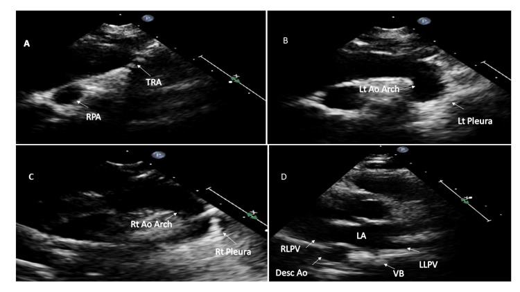
Figure 6. Sequential two-dimensional echocardiographic images of double aortic arch.
(A) Suprasternal long axis sweep with the probe position at 12 o'clock shows tracheal cartilages (TRA). (B) Tilting the probe leftwards shows the left aortic arch (Lt Ao Arch) and left pleura (Lt pleura). (C) Tilting the probe rightwards from 12 o'clock shows the right aortic arch (Rt Ao Arch) and right pleura (Rt pleura). (D) Right descending thoracic aorta (Desc Ao) in this patient with double aortic arch.
RPA: right pulmonary artery; VB: vertebral body; RLPV: right lower pulmonary vein; LLPV: left lower pulmonary vein; RPA: right pulmonary artery; LA: left atrium.

