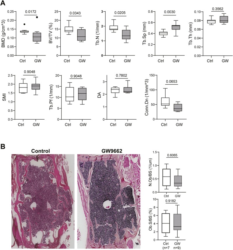
Figure 1.
(A) Microcomputed tomography (μCT) data showing bone mineral density (BMD) and bone structural parameters of vertebrae from 25-month-old female C57BL/6 mice treated without or with GW9662 for 6 weeks. Values are given as mean ± SD; n = 9–10 mice per group. Unpaired t test, p values are indicated. Symbols (dot and square, included in data set) denote outliers. BV/TV = bone volume/tissue volume; Tb.N = trabecular number; Tb.Th = trabecular thickness; Tb.Sp = trabecular spacing; SMI = structure model index, a parameter indicating the plate- or rod-like geometry of trabecular structures; Tb.Pf = trabecular pattern factor; DA = degree of anisotropy; Conn.Dn. = connectivity density. (B) Histology and histomorphometry analyses. Representative hematoxylin and eosin (H&E) stained vertebrae sections from 25-month-old female C57BL/6 mice treated without or with GW9662 for 6 weeks. Quantitative results for osteoblast number and osteoblast-covered bone surface area are shown in graphs. Data are shown as mean ± SD, n = 7–9 mice per group. Unpaired t test and p values are indicated.

