Fig. 4.

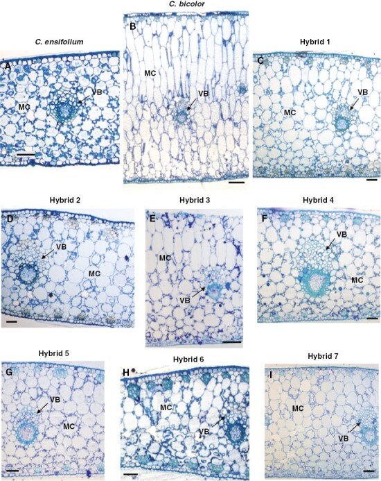
Inner structure of leaves of (A) C. ensifolium, (B) C. bicolor subsp. pubescens and (C–I) F1 hybrids 1–7. MC, mesophyll cell, VB, vascular bundle. Scale bars in (B) and (E) = 100 µm; in other panels = 50 µm.

Inner structure of leaves of (A) C. ensifolium, (B) C. bicolor subsp. pubescens and (C–I) F1 hybrids 1–7. MC, mesophyll cell, VB, vascular bundle. Scale bars in (B) and (E) = 100 µm; in other panels = 50 µm.