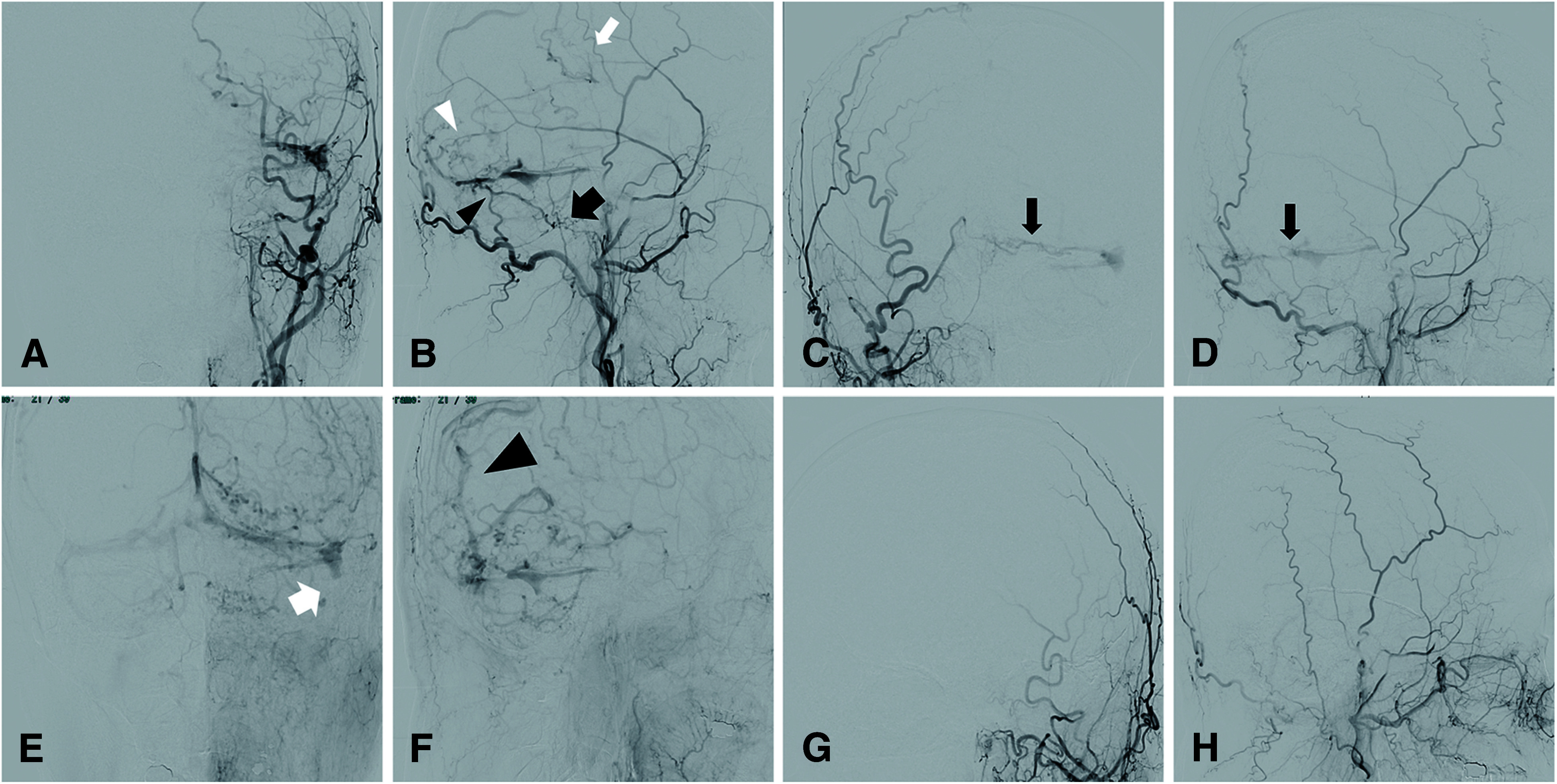
Fig. 3. DSA before and after transarterial embolizations. Left external carotid artery angiography demonstrates multiple arterial feeders to the transverse sinus. Arterial feeders include the OA (black arrowhead), ascending pharyngeal artery (large black arrow), and MMA (white arrowhead) (A and B). Furthermore, some feeders from MMA shunts directly to the cortical vein (B, white arrow). Right external carotid artery angiography demonstrates multiple arterial feeders from OA to the transverse sinus (C and D, black arrow). The proximal of left sigmoid sinus is occluded (E, large white arrow). The main outflow routes were through the confluence to the right transverse sinus and through the cortical vein to the SSS (F, large black arrowhead). Left external carotid artery angiography demonstrates no residual fistulas after endovascular treatments (G and H). MMA: middle meningeal artery; OA: occipital artery; SSS: superior sagittal sinus.

