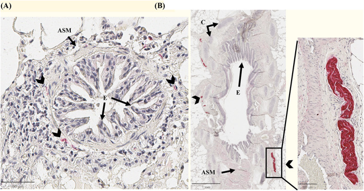
FIGURE 1.

Immunohistochemistry of peribronchial innervation. Representative images of the peribronchial innervation of a horse with asthma stained with anti‐s100 antibody (Vector Red, counterstained with hematoxylin). In panel A, small nerves (arrowheads) around a bronchiole. Panel B shows a large nerve in the surrounding of a bronchi. ASM, airway smooth muscle; C, cartilage; E, bronchi epithelium.
