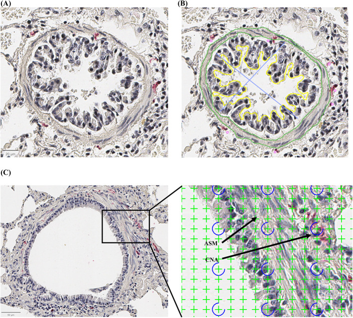
FIGURE 2.

Histomorphometric measurements by tracing and point counting. (A,B) Bronchiole (histological section, ×40). IP = internal perimeter (yellow), LA = lumen area (area inside the yellow outline), ID = internal diameters (blue lines), ASM = smooth muscle area (areas between the green lines), NBN = number of peribronchial nerves (pink dots). (C) Point counting technique using a grid of 1024 crosses per screen. Crosses were counted as either nerve (CNA, cumulative nerve area) if they encompassed red‐stained peribronchial tissue or as smooth muscle (ASM, smooth muscle area).
