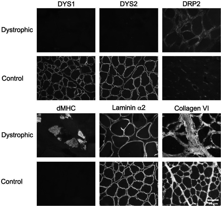
FIGURE 2.

Immunofluorescent staining of cryosections from the trapezius muscle of the dystrophic cat and archived control vastus lateralis muscle was performed for localization of dystrophy‐associated proteins. Antibodies against dystrophy‐associated proteins included those against the rod (DYS1) and carboxy terminus (DYS2) of dystrophin, utrophin (DRP2), developmental myosin heavy chain (dMHC) to demonstrate regenerating fibers, and against laminin α2 and collagen 6. Protein localization for both DYS1 and DYS2 could not be detected and utrophin was increased compared to archived control muscle. Regenerating fibers were highlighted by staining for dMHC. Staining for both laminin α2 and collagen 6 was like control tissue. Bar in lower right image equals 50 μm for all images.
