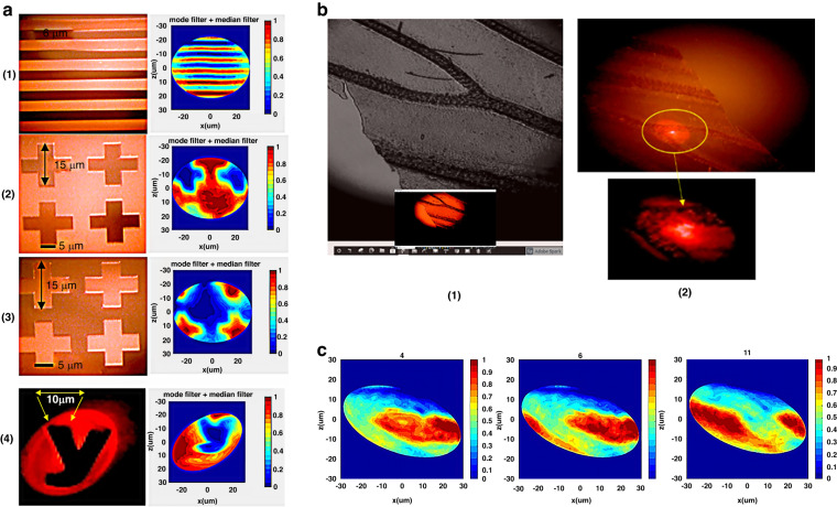
Fig. 7. Reconstructed image results.
a Masks (photographs left) and images reconstructed by the APD (right), (1) line pattern with line and gap widths of 5 µm, (2) solid cross pattern with 15 µm length and 5 µm gaps, (3) hollow cross pattern with 15 µm length and 5 µm gap, (4) letter “y” with 10 μm line width. b Psychodidae wing illumination. (1) Microscopy image of the wing, (2) area of the wing illuminated by the scanner. c Images captured by the APD, showing wing structures as the FOV moves from right to left

