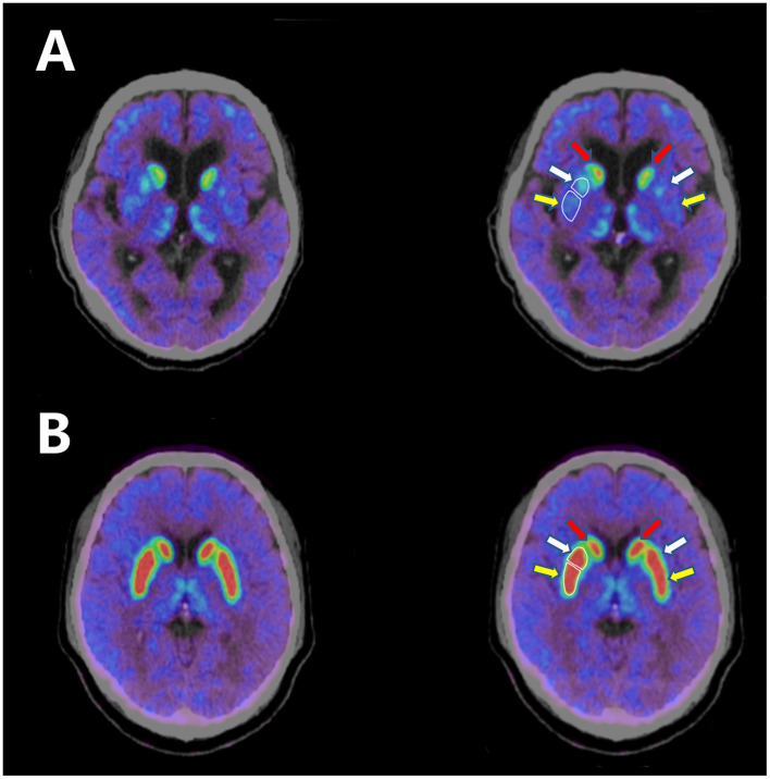
Figure 1.
(A) 18F-AV133 PET-CT scan of the patient shows significant decrease in uptake in the bilateral putamen. (B) 18F-AV133 PET-CT scan of a healthy person shows symmetrical uptake in both the bilateral caudate nucleus and the putamen. White enclosed lines distinguish between the anterior putamen and the posterior putamen. Red arrow: caudate nucleus; white arrow: anterior putamen; yellow arrow: posterior putamen.

