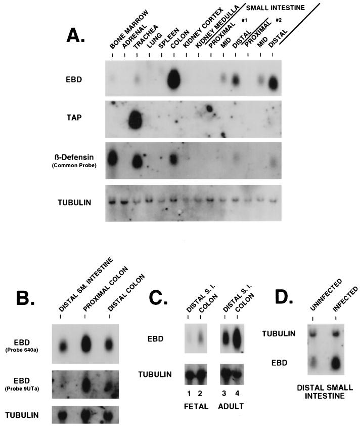
FIG. 4.
Northern blot analysis of EBD gene expression in bovine tissues. (A) Tissue distribution of β-defensins. Total RNA (20 μg) extracted from 14 different tissues was resolved by denaturing gel electrophoresis, capillary blotted to a nylon filter, and probed with either EBD 285a as an EBD probe, TAP286a as a TAP-specific probe, TAP48a as a common probe for β-defensins, or an α-tubulin probe. The hybridization and wash conditions were as described in Materials and Methods. Small intestine samples designated #1 and #2 represent RNAs extracted from the tissues of two healthy cows. (B) Expression of EBD in colonic tissue and usage of the putative upstream transcription start site. Total RNAs from the distal small (SM.) intestine, proximal colon (10 cm from the ileocecal junction), and distal colon (10 cm from the rectum) were analyzed as for panel A. The Northern blot was hybridized with EBD 285a to assess distribution of expression. The same blot was stripped of probe and rehybridized with EBD 9UTa, a probe from the unique sequence found in the 5′-extended RACE clone (Fig. 2, clone APT131.5) (see text). (C) Comparison of fetal and adult tissue expression of EBD. Total RNAs were isolated from the distal small intestine (S.I.) and colon of a bovine fetus at 4 months gestational age and from corresponding tissues of an adult cow and then analyzed as for panel A. (D) Northern blot analysis of EBD mRNA in enteric tissue from a calf infected with C. parvum. Total RNA was isolated from the distal 20 cm of small intestine from a C. parvum-infected calf and from a control uninfected calf. Analysis was as for panel A.

