
Figure 2. Design of hyperglycosylated trihead antigens from additional H1 HAs.
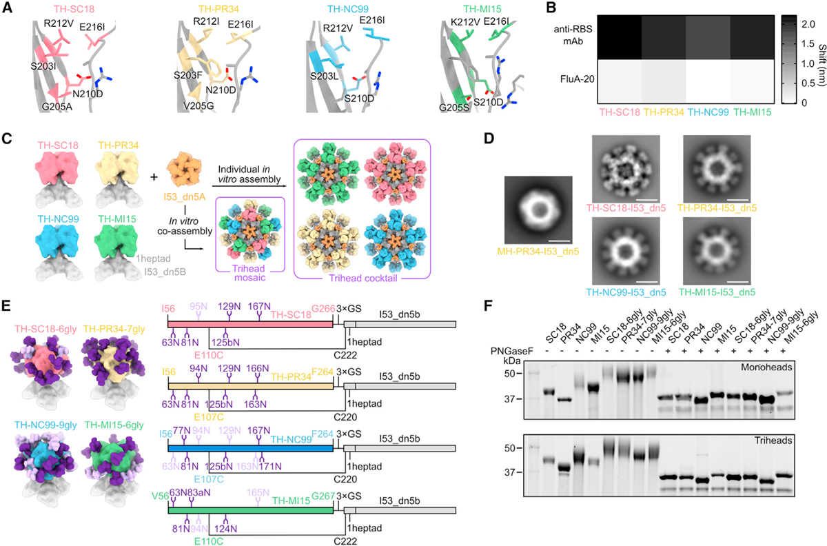
(A) Diagram of RBD trimer interfaces for TH-SC18, TH-PR34, TH-NC99, and TH-MI15, where mutated residues are colored and labeled.
(B) BLI of trihead components against RBS-directed mAbs (5J8, anti-PR34, and C05) and FluA-20.
(C) Schematic of TH-SC18, TH-PR34, TH-NC99, and TH-MI15 constructs and their in vitro assembly into mosaic or cocktail I53_dn5 nanoparticles.
(D) nsEM 2D class averages of MH-PR34-I53_dn5 and trihead I53_dn5 nanoparticles. Scale bars = 25 nm.
(E) Model structures and gene diagrams for hyperglycosylated triheads with wild-type glycans in light purple and glycan knockins in dark purple. Strain-specific H1 HA numbering is in respective HA strain color, and trihead model numbering is in black.
(F) Reducing SDS-PAGE of wild-type and hyperglycosylated monoheads and triheads without and with PNGaseF digestion.