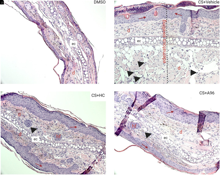
Fig. 5.
Histologic images of skin injuries after CS tear gas agent exposure and treatment with TRPA1 antagonists. Right ears of C57BL/6 male mice were exposed to CS (200 mM, 20 μl) and left ears to DMSO (solvent for CS, 20 μl). At 0.5, 4, 24, and 48 hours after CS exposure, mice were treated with vehicle (0.5% methylcellulose), HC-030031 (HC), or A967079 (A96) intraperitoneally (i.p.). Representative H&E-stained histopathologic sections from (A) DMSO, (B) CS + vehicle, (C) CS + HC-030031, and (D) CS + A967079 groups are presented at 20× magnification. Black arrow heads = infiltration of leukocytes; red arrow = epidermal thickening. d, dermis; e, epidermis; ec, elastic cartilage; hf = hair follicle.

