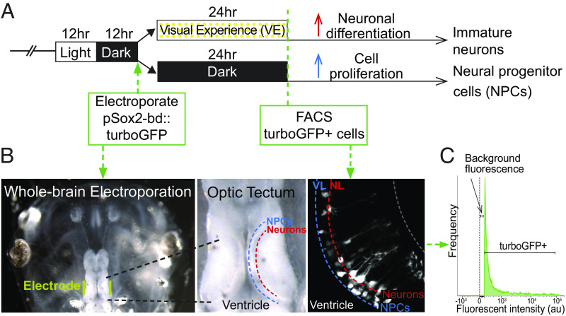
Fig. 1.
Isolation of neural progenitor cells and immature neurons from the optic tectum. (A) Visual experience paradigm used to enrich for NPCs and immature neurons. Animals were reared in 12 h light/12 h dark until stage 46 when the midbrain was electroporated with pSOX2-bd::tGFP plasmid. After electroporation, animals were randomly divided into two groups, one exposed to enhanced visual experience (VE) for 24 h. (B) Whole brain electroporation labels NPCs and neurons in the optic tectum. Left: Image of the head of the tadpole with electrodes (yellow) on each side of the optic tectum. Middle: Dorsal view of the optic tectum. The ventricle, NPC layer, and neuronal layer are labeled. Right: in vivo 2-photon image of tGFP-labeled NPCs and neurons in the ventricular layer (VL) and neuronal layer (NL). (C) Fluorescence histogram indicates the gate setting of the fluorescence-activated cell sorting (FACS) to isolate tGFP+ cells. Control is set to the background fluorescence from nonelectroporated midbrain cells.

