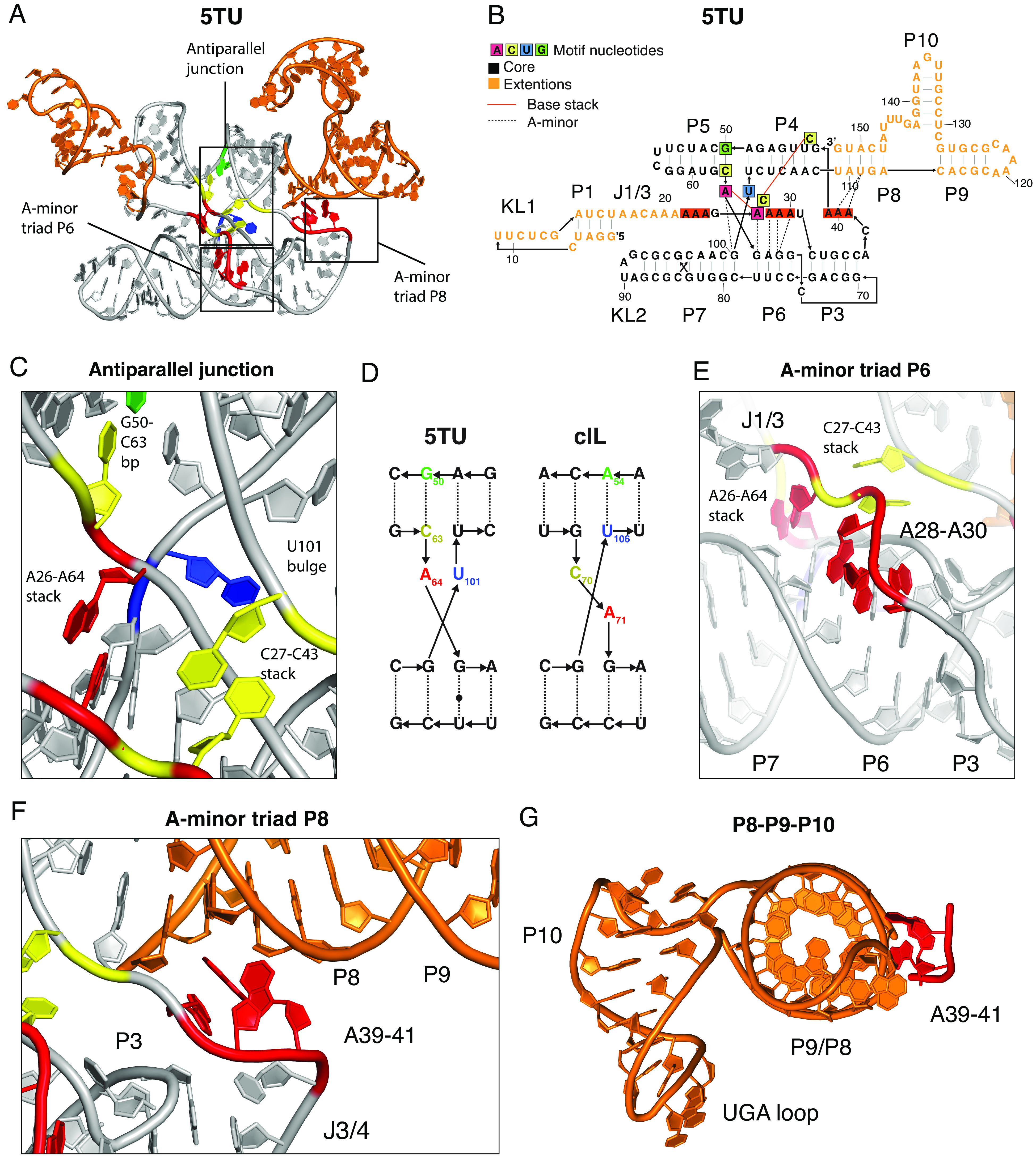
Fig. 4.

Structural features of catalytic subunit 5TU. (A) Atomic model of the 5TU subunit shown in cartoon representation with nucleotides in core motifs and extension regions colored as in panel B. Core motifs are indicated by boxes. (B) Secondary structure diagram of 5TU with annotation of tertiary interactions and core motif nucleotides. (C) Zoom on antiparallel junction and its interaction with J1/3 showing central base pairs, stacks, and bulges. (D) Diagram showing the changed junction between 5TU and its progenitor cIL. (E) Zoom on A-minor triad interacting with the minor groove of P6. Also shown is the A26-A64 stack that interacts with the minor groove of P6 and P7. Depth fog is used to highlight the motif. (F) Zoom on A-minor triad of J3/4 interacting with the minor groove of P8. (G) The P8-P10 domain is shown along the helical axis of P8/P9 to show the perpendicular orientation of the P10 helix exposing a 3-nucleotide UGA loop. A39-41 is shown to highlight stabilization from core domain.
