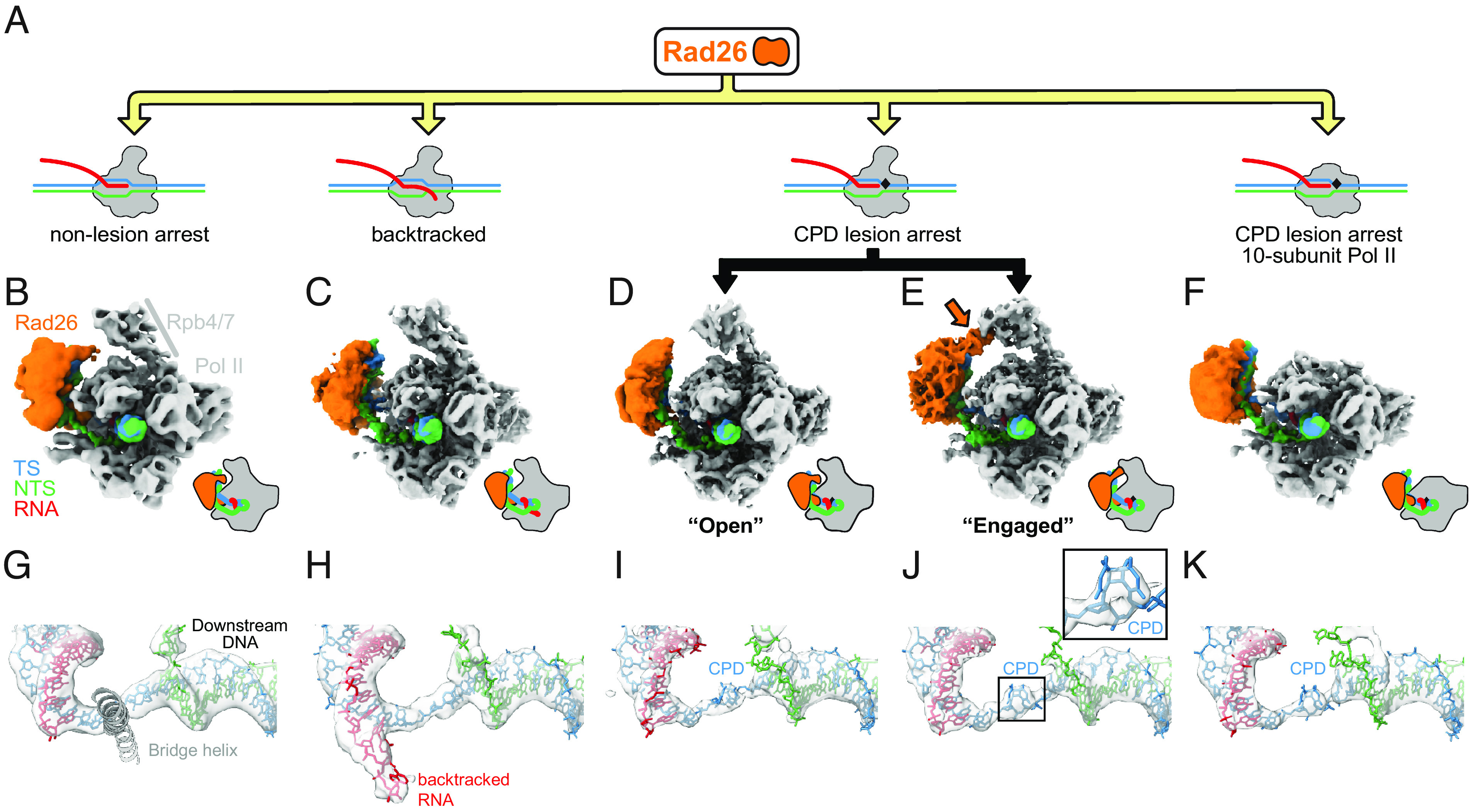
Fig. 1.

Rad26 interacts in a similar way with paused, CPD-stalled, and Backtracked RNA Pol II. (A) Cartoon representation of the different complexes analyzed by cryo-EM. The black rhomboid represents the CPD lesion. (B–F) Cryo-EM maps of (B) Pol II-Rad26 at a non-lesion arrest from our previous work (5.8 Å) (8), (C) Backtracked Pol II-Rad26 (4.4Å), (D, E) Pol II(CPD)-Rad26 in states showing either an “Open” (3.7 Å) (D), or “Engaged” (3.5 Å) (E) interaction between Rad26 and Rpb4/7 (orange arrow), and (F) Pol II(CPD)-Rad26 with Pol II lacking Rpb4/7 (4.7 Å). The maps were filtered according to the local resolution and were segmented and colored to highlight the different components, as indicated in (B). Cartoon representations of each structure, in the same orientation, are shown next to the maps. (G–K) Cryo-EM densities corresponding to the DNA/RNA scaffolds in the vicinity of the active site of Pol II segmented from the maps shown in (B–F). The active site Bridge helix was included as a reference point. A close-up of the cryo-EM density corresponding to the CPD lesion is shown in (J). The color scheme used throughout the paper is as follows: Pol II: gray; Rad26: orange; non-template strand: green; template strand: blue; RNA: red.
