Fig. 1.
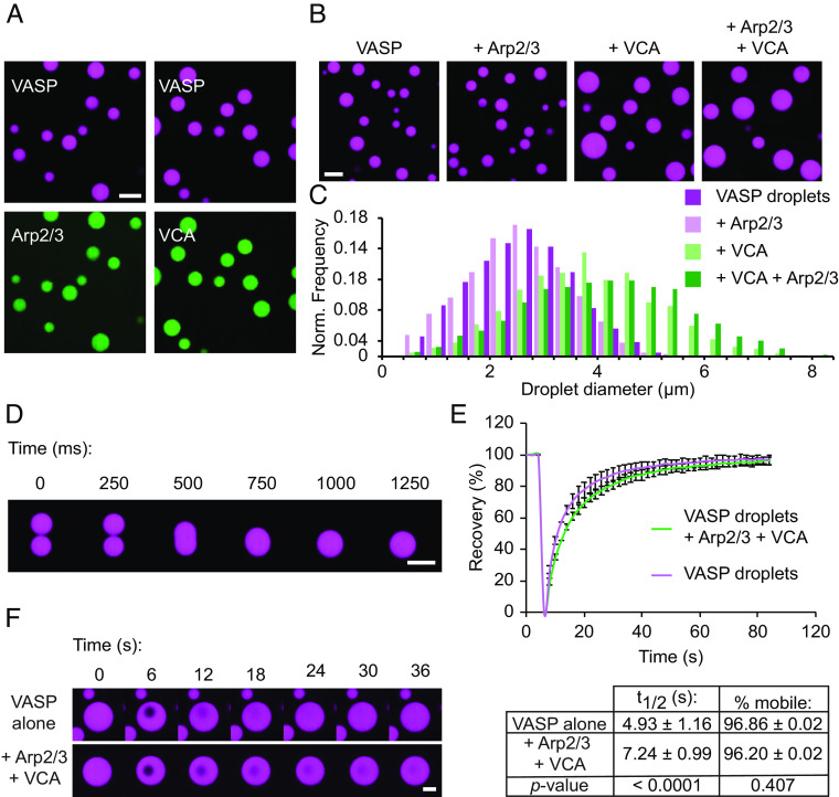
Arp2/3 and VCA participate in phase separation of liquid-like VASP droplets. (A) Enrichment of 1 μM Arp2/3 (labeled with Atto-594, shown in green) or 1 μM VCA (labeled with Atto-488, shown in green) to 20 μM VASP droplets. Droplets are formed in “droplet buffer”: 50 mM Tris pH 7.4, 150 mM NaCl, 3% (w/v) PEG8000. (Scale bar, 5 μm.) (B) Representative images of droplets formed from a 20 μM solution of VASP with either 150 nM Arp2/3, 6 μM VCA, or both 150 nM Arp2/3 and 6 μM VCA. (Scale bar, 5 μm.) (C) Histogram of droplet sizes quantified from B. Data from n = 3 replicates. (D) Composite droplets formed of 20 μM VASP, 150 nM Arp2/3, and 6 μM VCA still remain liquid-like, as exhibited by rapid fusion events. (Scale bar, 5 μm.) (E) Composite droplets formed of 20 μM VASP, 150 nM Arp2/3, and 6 μM VCA display rapid recovery after photobleaching, and the fraction of mobile VASP is nearly equivalent to droplets lacking Arp2/3 and VCA. Curves show the average VASP recovery profile with error bars representing SD for n = 13 droplets for each condition. Inset table shows t1/2 and percent mobile fraction for each condition, with corresponding P-values from an unpaired two-tailed t-test. (F) Representative time series of FRAP of either VASP-only droplets, or composite droplets as quantified in E. (Scale bar, 2 μm.)

