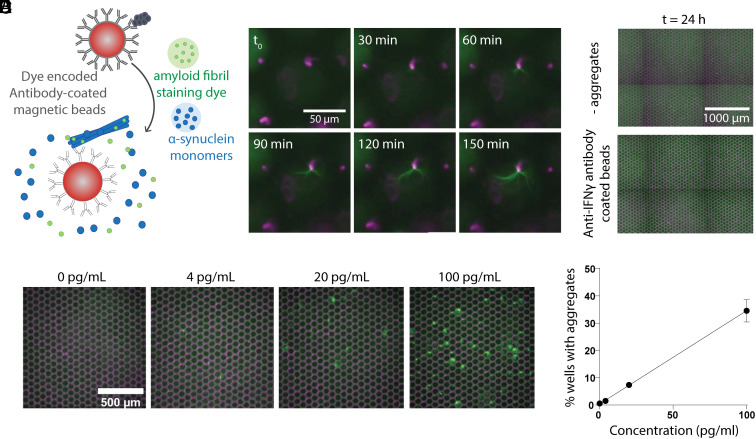
Fig. 4.
Bead-based digital SAA. (A) Schematic depicting the assay where aggregates are first captured on antibody-coated magnetic beads and then combined with SAA reaction components before microwell loading. (B) Images showing filament growth from a single bead over the course of 150 min. (C) Representative images after 24 h of running the bead-based assay when the beads were incubated without pre-formed filaments (− aggregates) and when pre-formed filaments were incubated with non-specific antibody-coated beads (anti-IFNγ). (D) Microwell images 24 h after incubating the bead-based SAA at varying concentrations of pre-captured pre-formed filaments. (E) The percent of wells with growing aggregates vs. the concentration of pre-formed fibrils incubated with antibody-coated beads.

