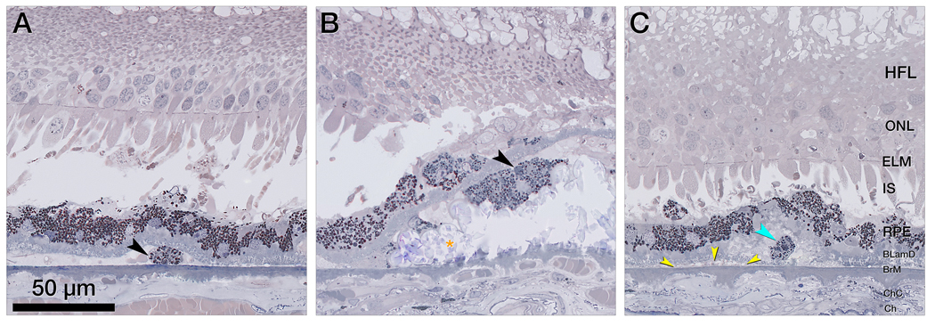
Figure 6. Retinal pigment epithelium (RPE)-originated subducted cells and granule aggregates, under the RPE cell layer.
A&B. Magnified histology shows subducted retinal pigment epithelium (RPE) cells in the sub RPE- basal lamina (BL) space, with distinct content of lipofuscin and melanolipofuscin granules. Photoreceptors are short and mostly lack outer segments, Bruch’s membrane (BrM) is intact, and the choriocapillaris (ChC) is atrophic. A. In a single subducted RPE cell (black arrowhead), granule packing density is slightly lower than in-layer RPE. B. A cluster of subducted cells (black arrowhead) adjacent to a calcified nodules (orange asterisk) shows granule packing density and reflectivity like in-layer RPE, which is in turn less than the cells in panel A. The subducted cells are separated from the in-layer cells by basal laminar deposit (BLamD). C. Subducted cells are distinct from the shedding RPE phenotype, which releases granule aggregates (teal arrowhead) into underlying BLamD; here, within a basal mound (soft drusen material). It is bounded by the native RPE-basal lamina and is therefore separate from basal linear deposit in sub-RPE-BL space (yellow arrowheads).
BLamD, basal laminar deposit; BrM, Bruch’s membrane; Ch, choroid; ChC, choriocapillaris; ELM, external limiting membrane; HFL, Henle fiber layer; IS, inner segments; NIR, near infrared reflectance; OCT, optical coherence tomography; ONL, outer nuclear layer; RPE, retinal pigment epithelium sub-RPE-BL, subretinal pigment epithelium basal lamina.

