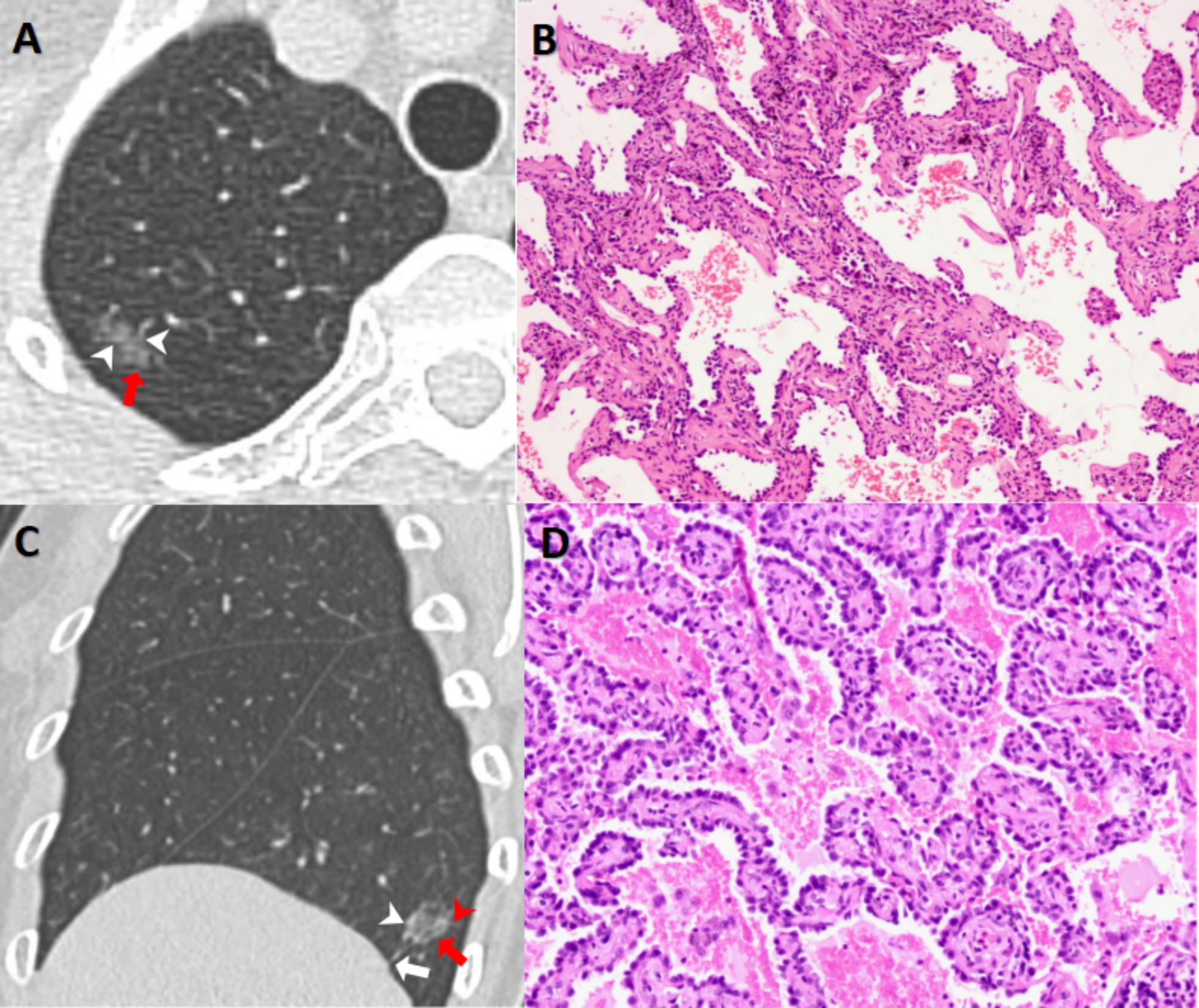
Fig. 3.

GGNs with different pathological types. (A&B, MIA) A GGN with an average diameter of 9.5 mm could be seen in the upper lobe of right lung (A, red arrows)in a 71-year-old female, and vascular shadows (A, white arrowheads) could be seen inside it. Its score of Lung-RADS and PNI-GARS were 2 and IIIb,respectively.The final pathological diagnosis (B, HE×200) was microinvasive adenocarcinoma (MIA). (C&D, IAC) A GGN with a mean diameter of 11.4 mm could be seen in the lower lobe of right lung (C, red arrows) in a 56-year-old female, and vessels (C, white arrowheads), spiculation (C, red arrowheads ) and pleural indentation signs (C, white arrows) could be seen. Its score of Lung-RADS and PNI-GARS were 2 and IV, respectively. The final pathological diagnosis (D, HE×400) was invasive adenocarcinoma (IAC)
