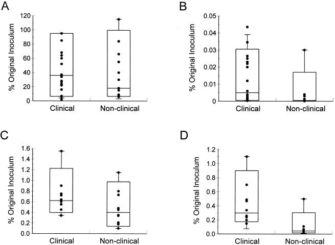
FIG. 1.
Abilities of clinical and nonclinical strains of Y. enterocolitica biotype 1A to adhere to (A) and invade (B) HEp-2 cells and to adhere to (C) and invade (D) CHO cells. Approximately 107 CFU was incubated with epithelial cells for 3 h before nonadherent bacteria were removed by three washes. To determine the number of adhesive bacteria, some epithelial cells were lysed and the cell-associated bacteria were enumerated. To determine the number of intracellular bacteria, other epithelial cells were incubated in fresh tissue culture medium containing 100 μg of gentamicin/ml for 90 min before epithelial cells were lysed and bacteria were enumerated. Data are expressed as percentages of the original inoculum and are means from at least two separate experiments using duplicate wells. In the box-and-whisker plots, the horizontal line within the box is the median value, the limits of the box are the 10th and 90th percentiles, and the whiskers are the 5th and 95th percentiles. The difference in adhesion between the two groups of isolates is not significant (P = 0.3 for HEp-2 cells and P = 0.14 for CHO cells by the Mann-Whitney test). The difference in invasion between the two groups of isolates is significant (P = 0.002 for HEp-2 cells and P < 0.001 for CHO cells).

