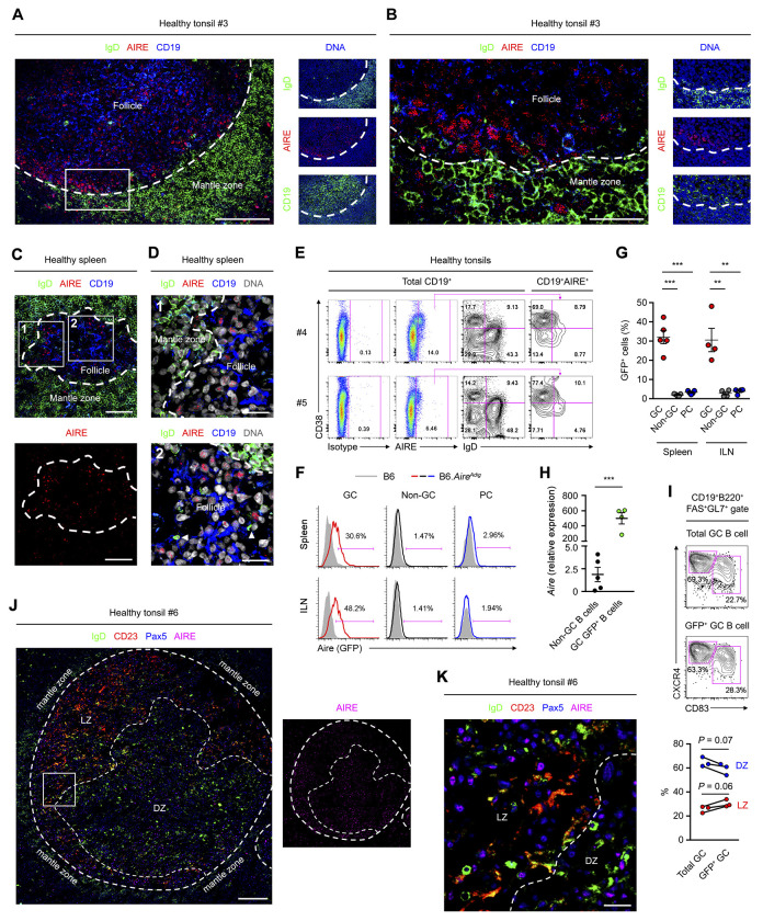
Figure 1. GC B Cells Express AIRE.
(A and B) Immunofluorescence analysis of the tonsillar tissue of a healthy donor for IgD, CD19, AIRE and DAPI-stained DNA. The dotted line outlines the follicles. Bars: 100 μm (A) or 25 μm (B).
(C and D) Immunofluorescence analysis of tissues of a healthy donor for IgD, AIRE, CD19 and DNA. Arrow heads indicate follicular IgD+ plasmablasts. Dotted lines mark the boundary between follicular mantle zone and the follicle. Bars: 40 μm (C) and 15 μm (D).
(E) Flow cytometric analysis of AIRE expression in tonsillar total CD19+ B cells, IgD+CD38− naive B cells, IgD+CD38+ founder GC (FGC) B cells, IgD−CD38+ GC B cells and IgD−CD38− memory B cells. The data represent 5 donors.
(F and G) Flow cytometric and statistical analyses of Aire (GFP) expression in splenic and ILN viable CD19+B220+FAS+GL7+ GC B cells, CD19+B220+FAS−GL7− non-GC B cells and CD19loB220loCD138+ PCs of B6 mice (shaded histograms, n = 5 for spleen and n = 4 for ILN) or B6.AireAdig mice (colored histograms, n = 5 for spleen and n = 4 for ILN) after 4 i.p. immunizations with NP32-KLH with CFA and subsequently with IFA. Data are represented as mean ± SEM. **P < 0.01, ***P < 0.001, by 1-way ANOVA with Tukey’s post hoc test.
(H) qPCR analysis of Aire transcript levels in CD19+B220+FAS−GL7− non-GC B cells (n = 5) and CD19+B220+FAS+GL7+GFP+ Aire-expressing GC B cells (n = 4) of AireAdig mice after 1 i.p. immunization with SRBC and CFA. Data are represented as mean ± SEM. ***P < 0.001, by 2-tailed unpaired t-test.
(I) Flow cytometric and statistical analyses of CD83+CXCR4lo LZ and CD83−CXCR4hi DZ B cells in splenic total GC and GFP+ GC B cells of immunized B6.AireAdig mice, by 2-tailed paired t-test.
(J and K) Immunofluorescence analysis of the tonsillar tissue of a healthy donor for IgD, CD23, Pax5 and AIRE. The dotted line outlines the follicles and delineates the border between LZ and DZ. Bars: 200 μm (J) or 30 μm (K).

