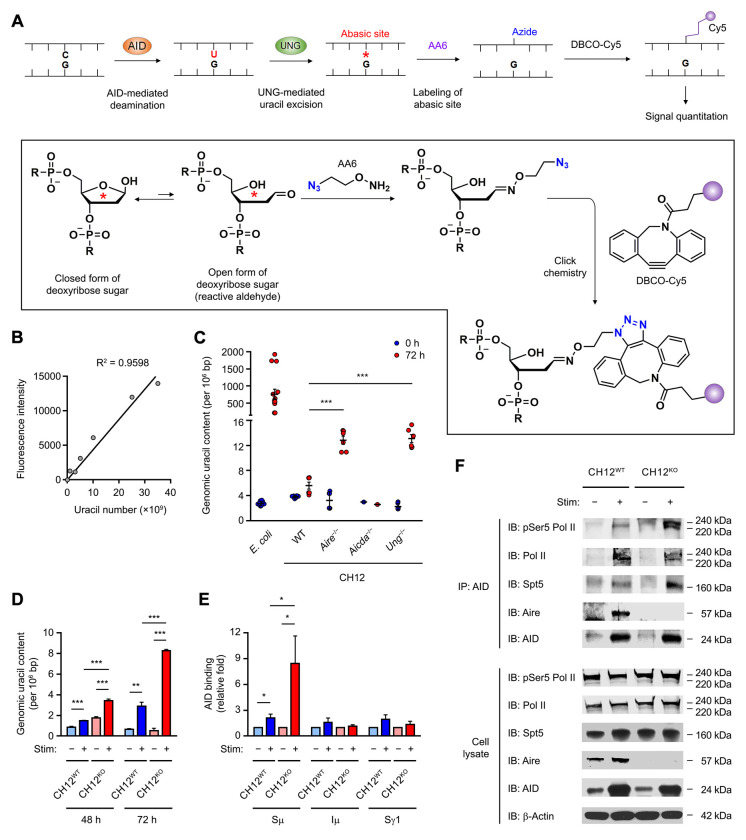
Figure 6. AIRE Inhibits AID Function by Interfering with AID Targeting to Its IgH DNA Substrate.
(A and B) The principle, chemistry and calibration of the dot blot assay for the quantitation of genomic uracil content.
(C) The genomic uracil levels in WT, Aire−/−, Aicda−/− or Ung−/− CH12 cells after 72 h of treatment without or with anti-CD40, TGF-β and IL-4. The results are presented as mean ± SEM and represent 3 experiments. ***P < 0.001, by 2-tailed unpaired t-test. Bisulfite-treated E. coli DNA was included as a positive control.
(D) The genomic uracil content in WT and Aire−/− CH12 cells after 48 or 72 h of treatment without or with anti-CD40, TGF-β and IL-4. The results are presented as mean ± SEM and represent 3 experiments. **P < 0.01, ***P < 0.001, by 2-tailed unpaired t-test.
(E) ChIP-qPCR analysis for the interaction of AID with Sμ, Iμ and Sγ1 regions in WT and Aire−/− CH12 cells after 72 h of treatment without or with anti-CD40, TGF-β and IL-4. The results are presented as mean ± SEM and represent 3 experiments. *P < 0.05, by 2-tailed unpaired t-test.
(F) Co-IP of AID with pSer5-Pol II, total Pol II, Spt5 and Aire in WT and Aire−/− CH12 cells after 72 h of treatment without or with anti-CD40, TGF-β and IL-4. The results represent 3 experiments.

