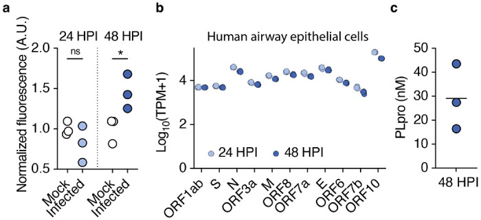
Figure 1. SARS-CoV-2 PLpro is released from infected airway epithelial cells.

a, PLpro proteolytic activity from the supernatant of SARS-CoV-2 or mock infected Calu-3 cells 24 and 48 HPI, measured by the fluorescence of cleaved substrate, normalized across samples from the same day. One-way ANOVA: p=0.014, F(3,8)=6.790; Holm-šídák’s multiple comparisons, pmock vs. infected 24h =0.244, pmock vs. infected 48h =0.030, n=3 wells per group. b, SARS-CoV-2 transcripts from Calu-3 cells infected with SARS-CoV-2 (multiplicity of infection = 1) for 24 and 48 hours post infection (HPI), transcripts per million (TPM), n=3 wells. c, Interpolated concentration of PLpro in the supernatant of Calu-3 cells 48 hours post infection, line represents mean.
