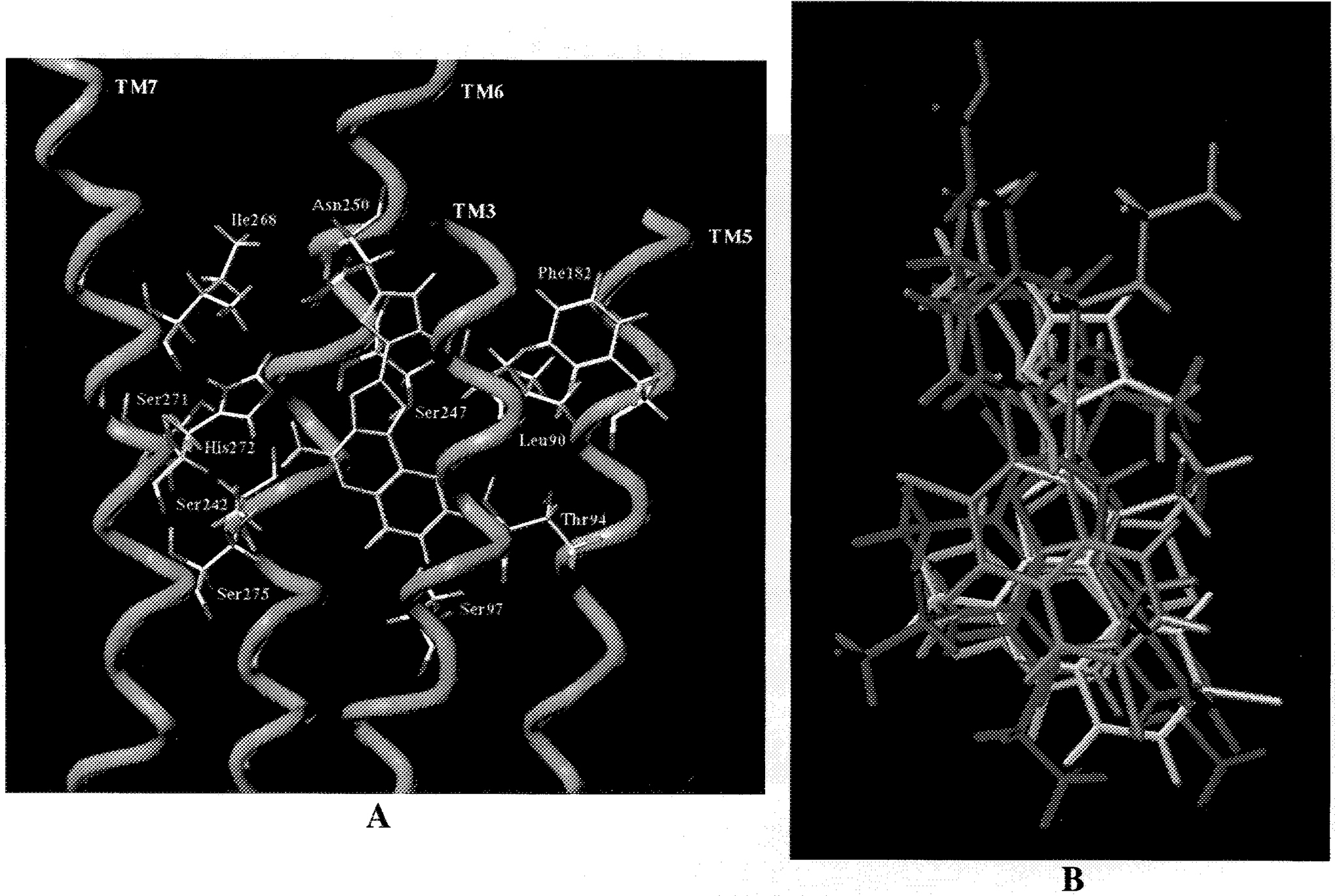
Figure 5.

(A, left) Side view of the A3–CGS 15943 complex model. The side chains of the important residues in proximity to the docked CGS 15943 molecule are highlighted and labeled. Residues in proximity (≤5 Å) to the docked CGS15943 molecule: Leu90 (TM3), Phe182 (TM5), Ser242 (TM6), Ser247 (TM6), Asn250 (TM6), Ser271 (TM7), His272 (TM7), Ser275 (TM7). (B, right) Superposition of all docked A3 antagonist structures. The transmembrane helical bundle is not highlighted, but it conserves the same arrangement shown in Figure 5A. Blue = MRS 1476; green = L-268,605; yellow = CGS 15943; magenta = MRS 1067; orange = HE-EA.
