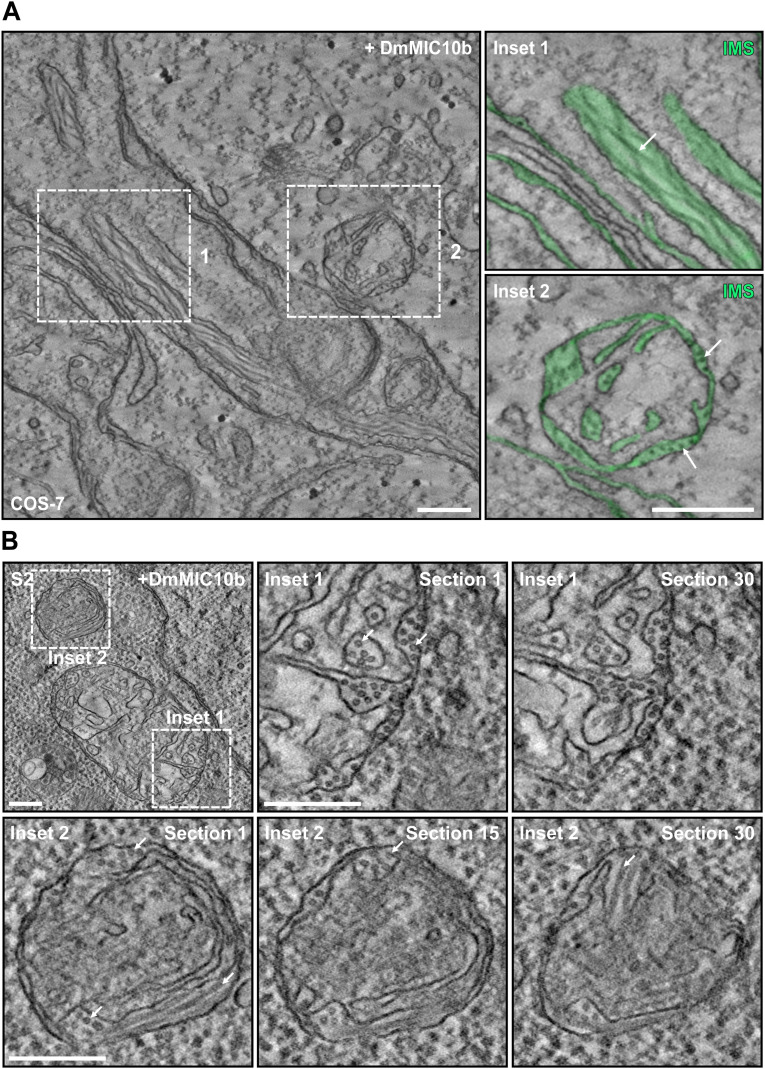
Figure S8. Localization of DmMIC10b filaments in mitochondria of COS-7 and S2 cells.
(A) Electron tomography of mitochondria from COS-7 cells expressing DmMIC10b-FLAG. The IMS, including the crista lumen, is colored in green. Arrows point to DmMIC10b-FLAG filaments located within the crista lumen (upper) and between OM and IBM (lower). (B) S2 cells transiently expressing DmMIC10b-FLAG were chemically fixed and analyzed by electron tomography. Shown are different sections of the two areas indicated by the dashed boxes. Arrows indicate filaments placed between the OM and the IBM and inside the crista lumen. Scale bars: 250 nm (A) and 200 nm (B).

