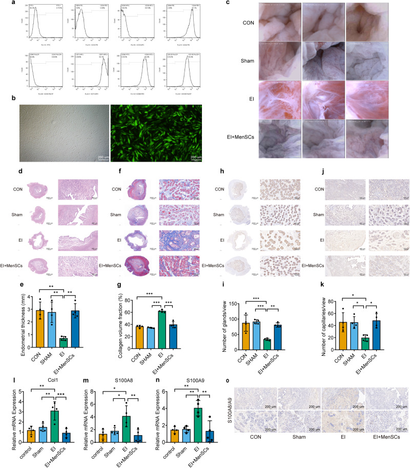
Fig. 8. MenSCs treatment promotes the repair of endometrial injury in a porcine model of IUA.
a Flow cytometry analysis of surface markers of MenSCs. b Morphology of MenSCs under light microscope and fluorescence microscope (green), scale bar = 200 μm. c Endometrium observed by hysteroscopy on postoperative day 35. d, e HE staining of porcine endometrial tissue sections, measuring endometrial thickness, n = 4 per group, ordinary one-way ANOVA with Tukey’s multiple comparison test. f, g Masson staining of porcine endometrial tissue sections, measuring collagen volume fraction, n = 4 per group, ordinary one-way ANOVA with Tukey’s multiple comparison test. h, i CK18 staining of porcine endometrial tissue sections, measuring glandular density, n = 4 per group, ordinary one-way ANOVA with Tukey’s multiple comparison test. j, k vWF staining of porcine endometrial tissue sections, measuring capillaries density, n = 4 per group, ordinary one-way ANOVA with Tukey’s multiple comparison test. l–n RT-PCR detection of Col1, S100A8, and S100A9 mRNA levels in each group, n = 4 per group, ordinary one-way ANOVA with Tukey’s multiple comparison test. o Staining results for S100A8/A9 in porcine endometrial tissues, scale bar = 200 μm. Data are presented as means ± SD. *P < 0.05, **P < 0.01, ***P < 0.001 compared with indicated groups.

