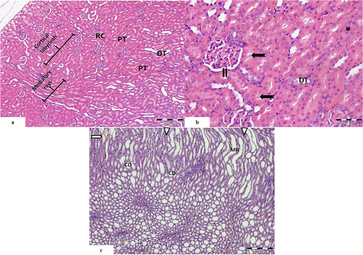
Fig. 1.
Light photomicrographs of rat’s kidney control group showing a renal cortex arranged into cortical labyrinths and medullary rays. It shows normal appearance of the renal corpuscle (RC), proximal convoluted tubules (PT), and distal convoluted tubules (DT). b Higher magnification showing the Bowman’s space of the renal corpuscle (
 ). Proximal convoluted tubules showing prominent brush border (
 ). The distal convoluted tubules (DT) appear with wider lumen and cuboidal cells with rounded nuclei. c The medulla is seen occupied by medullary rays (MR), collecting ducts (CD), renal interstitium (
 ), and blood vessels (
 ). (H&E. Mic.Mag a ×100, b ×400, and c ×100)

