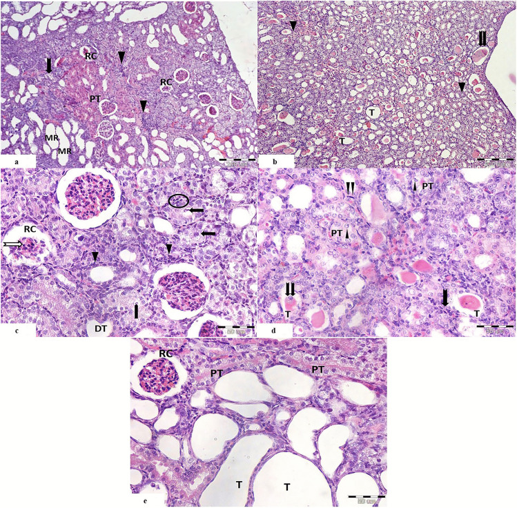
Fig. 2.
Light photomicrographs of a section of renal cortex from cisplatin-treated group showing a the cortex with distorted renal corpuscles (RC), ballooning of the tubules of the medullary rays (MR), intense cellular infiltration (
 ) among the degenerated tubules (
 ), and focal areas of proximal tubules with acidophilic cytoplasm (PT). b The medulla shows distorted tubules with multiple hyaline casts within their lumina (
 ). Some of the tubules appears dilated (T), and others show obliterated lumen (
 ). c The renal corpuscles (RC) appear with shrunken glomerular tuft and pyknotic nuclei (
 ). Peritubular cellular infiltration is noticed around the distorted tubules (
 ). The proximal convoluted tubules show vaculations (
 ), bizzare shaped nuclei of the lining cells (
 ), and extruded cells in the lumen with loss of brush border and basophilic cytoplasm (
 ). Distorted distal convoluted tubules with flattened cells are seen (DT). d The proximal tubules (PT) show karyolitic nuclei (
 ). Some tubules (T) show hyaline casts and exfoliated cells within their lumina (
 ). There are peritubular cellular infiltration (
 ) and proliferating interstitial fibroblasts (
 ). e Renal corpuscle (RC) appears with shrunken glomerular tuft and dark nuclei. Many tubules show severe ballooning (T). Cells of proximal convoluted tubules show vaculations with loss of brush boarder (PT). (H&E. Mic.Mag a, b ×100, c–e ×400)

