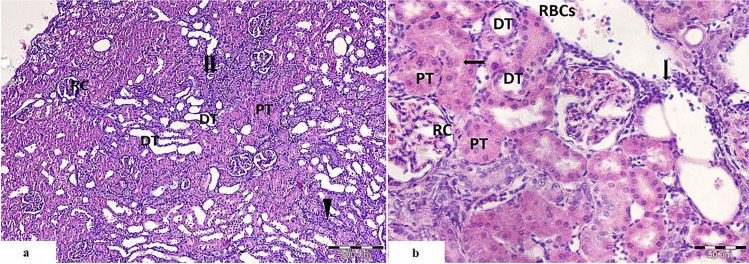
Fig. 3.
Light photomicrographs of rat’s kidney from cisplatin and PRP-treated group showing a, b the renal corpuscles appear nearly normal with bifurcated glomerular tuft (RC), many normal proximal convoluted tubules with eosinophilic cytoplasm and intact epithelial lining (PT). Few tubules are obliterated, with basophilic cytoplasm (
 ), and others appeared degenerated with hyalinized material in their lumen (
 ). b Distal convoluted tubules appear with cuboidal epithelial lining (DT). Widened interstitial space with evident cellular infiltration (
 ) and extravasated RBCs (RBCs) are also seen (
 ); brush border of PCT. (H&E. Mic.Mag a ×100 and b ×400)

