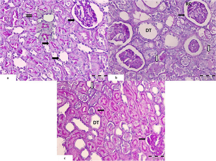
Fig. 7.
Light photomicrographs of renal cortex revealing a control group illustrates positive PAS reaction in the glomerulus, the basement membrane of the renal tubules (
 ), and brush border of proximal convoluted tubules (
 ). It is stained magenta red with the PAS stain. b The cisplatin-treated group shows thickened basement membrane of the glomerular tuft (
 ). One of the glomeruli shows hyper cellularity (RC). Many proximal convoluted tubules appear degenerated with thickened basement membrane around them (
 ). Some tubules show hyaline casts within its lumen (
 ). Some distal tubules show dilatation (DT). c Cisplatin and PRP-treated group shows positive reaction within the basement membrane of the renal corpuscle and the proximal convoluted tubules (
 ). Few of them appear degenerated with thickened basement membrane (
 ). Some distal tubules show dilatation with thickened epithelial basement membrane (DT), and others appear nearly normal (PAS. Mic.Mag ×400)

