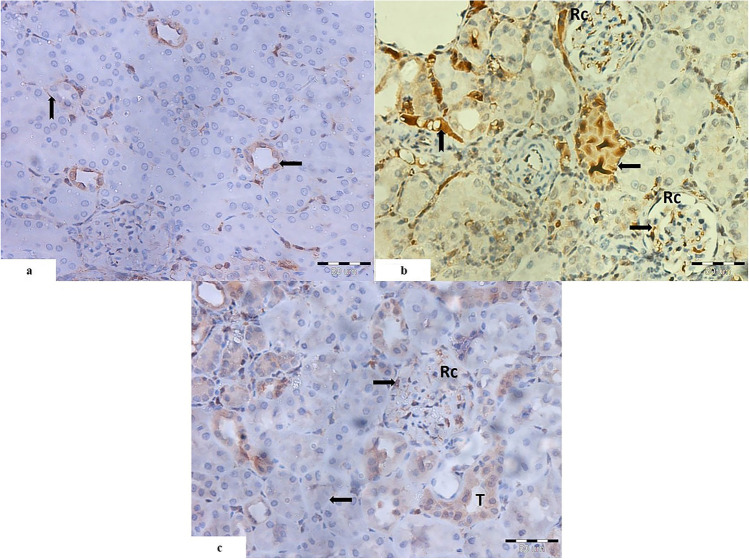
Fig. 8.
Light photomicrographs of section of the renal cortex: a the control group shows weak immunohistochemical reaction within the cells of the distal renal tubules (
 ) and the interstitium (
 ). b Cisplatin-treated group shows intense positive reaction in the degenerated tubules with obliterated lumen (
 ). The interstitium shows strong positive reaction (
 ). Also, there is strong positive reaction within the cells of the glomerular capillary tuft (
 ) of the renal corpuscle (RC). c Cisplatin and PRP-treated group shows positive reaction within the lining cells of some tubules (T), and others show weak reaction (
 ). Few cells of the glomerular tuft show positive reaction (
 ) (caspase-3 immunohistochemical staining. Mic.Mag ×400)

