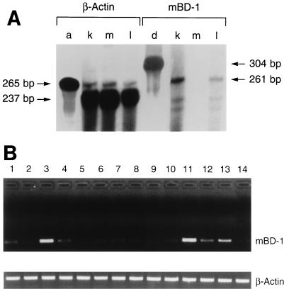
FIG. 5.
Analysis of mBD-1 expression by RNase protection analysis and RT-PCR (A) Measurement of mBD-1 expression by RNase protection analysis. Total RNA from the kidney (k), skeletal muscle (m), and whole lung (i.e., lung and trachea) (l) was investigated. Hybridization to a labeled riboprobe against β-actin (a) and mBD-1 (d) transcripts was performed a separate tube. (B) Detection of mBD-1 expression in various mouse tissues by nested RT-PCR. Poly(A)+ RNA was isolated from mouse tissues and reverse transcribed, and the cDNAs were amplified by using two pairs of mBD-1-specific primers. A single 250-bp band was amplified by using gene-specific primers. As a control, β-actin cDNA was amplified by using specific primers. Lanes: 1, trachea, 2, lung (lung parenchyma without cartilaginous airways); 3, tongue; 4, esophagus; 5, small bowel; 6, large bowel; 7, gall bladder; 8, pancreas; 9, skeletal muscle; 10, heart; 11, fallopian tube; 12, ovary; 13, vagina; 14, brain.

