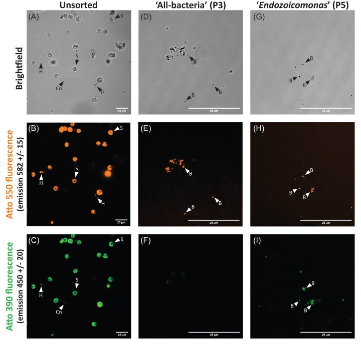
Figure 3.
A labelled A. loripes sample (colony Al13) prior to sorting with FACS (A–C) or sorted cell populations that were single (‘all-bacteria’, D–F), or dual (‘Endozoicomonas’, G–I) labelled. Each sample was visualized on a Nikon A1R confocal laser scanning microscope with channels for brightfield (A, D, G), 561 nm excitation (B, E, H), and 405 nm excitation (C, F, I). S=Symbiodiniaceae, Cn=host cnidocyst, H=uncharacterised host cell, B=bacteria. All scale bars are 25 µm.

