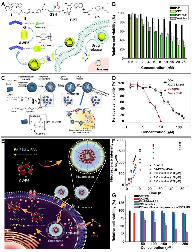
Figure 12.
(A) Schematic illustration of the formation of nanovesicle and its internalization progress; (B) Cell viability of MCF-7 cells after incubation with Cb, CPT, Cb + CPT mixture, and vesicles for 24 h. This figure is quoted with permission from Shao et al.[167]; (C) Schematic illustration of water-solution pillar[6]arene nanosponges (NS) in overcoming MDR; (D) Cell viability of MCF-7/ADR cells after incubation with DOX and DOX@NS. This figure is quoted with permission from Liu et al.[168]; (E) Schematic illustration of the preparation of PIC micelles and their application in inhibiting drug efflux; (F) Changes of extracellular fluorescence intensity after incubating with FA-PEG-b-PAA and different concentrations of PIC micelles; (G) Cell viability of MCF-7/ADR cells after incubation with different treatments. This figure is quoted with permission from Yu et al.[170]. AWP6: Anionic WP6; CPT: camptothecin; DOX: doxorubicin; FA: folic acid; GSH: glutathione; MDR: multidrug resistance; NS: nanosponge; PEG: poly (ethylene glycol); PIC: polyion complex.

