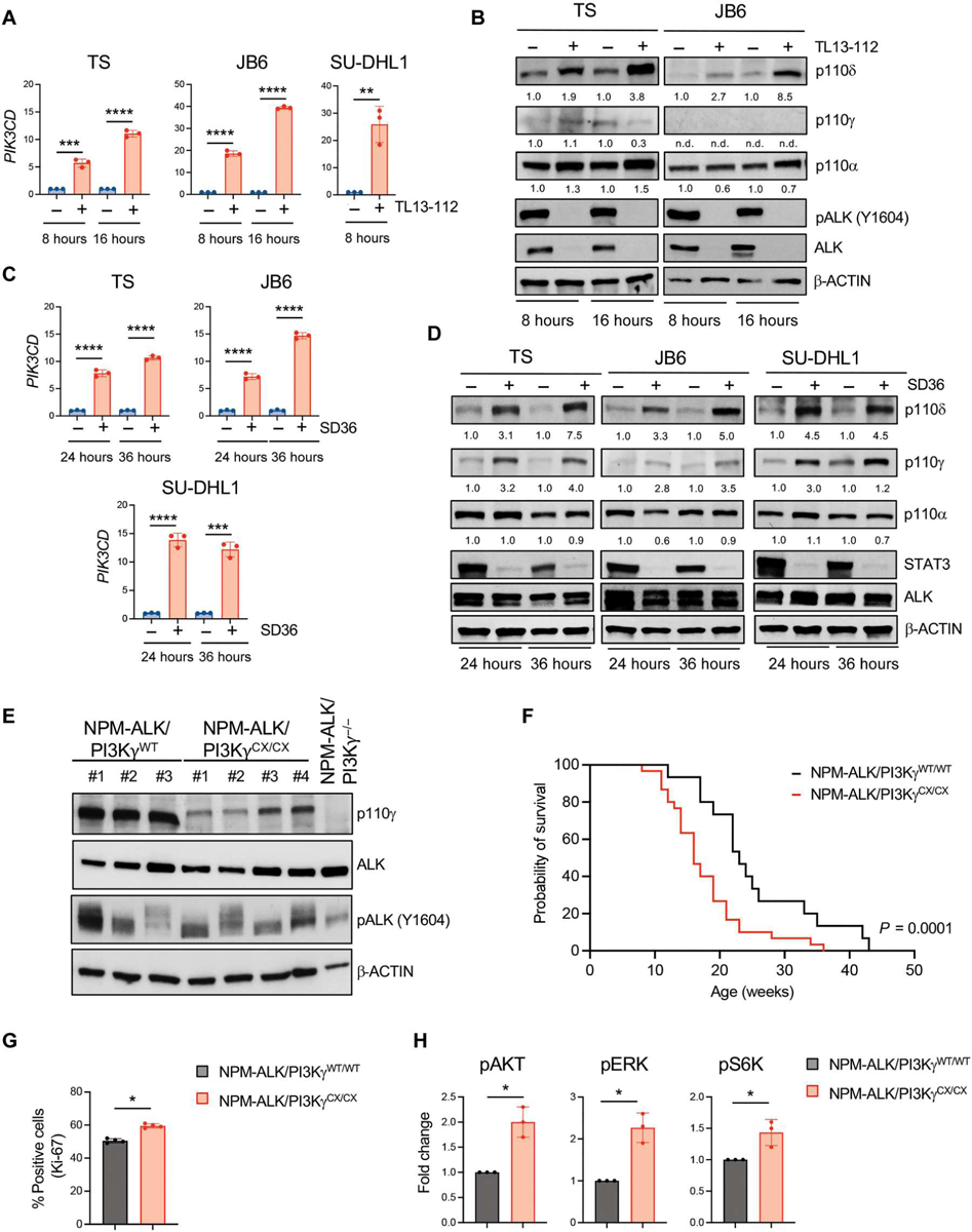
Fig. 3. PI3Kδ is repressed by ALK in ALK+ ALCL cell lines and constitutive activation of PI3Kγ accelerates ALK-dependent lymphomagenesis.

(A) qRT-PCR analysis of PIK3CD mRNA expression performed on TS, JB6, and SU-DHL1 cell lines treated with TL134–112 (100 nM). n = 3 technical replicates. (B) Western blot analysis on TS and JB6 cell lines treated with TL13–112 (100 nM). (C) qRT-PCR analysis of PIK3CD mRNA expression performed on ALK+ ALCL cell lines treated with SD36 (1 μM). n = 3 technical replicates. (D) Western blot analysis on ALK+ ALCL human cell lines treated with SD36 (1 μM). (E) Western blot analysis of lymphomas obtained from C57BL/6 mice with the indicated genotypes. (F) Kaplan-Meier survival analysis of NPM-ALK transgenic mice crossed with mice expressing an active form of PI3Kγ (PI3KγCX/CX) (black, NPM-ALK/PI3KγWT/WT, n = 30 mice; red, NPM-ALK/PI3KγCX/CX, n = 30 mice). ****P < 0.0001. Significance was determined by log-rank (Mantel-Cox) test. (G) Quantification of Ki-67–positive cells in sections of primary lymphomas (n = 4) with the indicated genotypes. (H) Amount of phosphorylated Akt, ERK, and S6K measured in murine primary tumors with the indicated genotypes by Bio-Rad Bio-Plex. *P < 0.05, **P < 0.01, ***P < 0.001, and ****P < 0.0001. Significance was determined by unpaired, two-tailed Student’s t test. Data are shown as means ± SD. For Western blots, β-actin was used as a loading control, and two independent experiments with similar results were performed.
