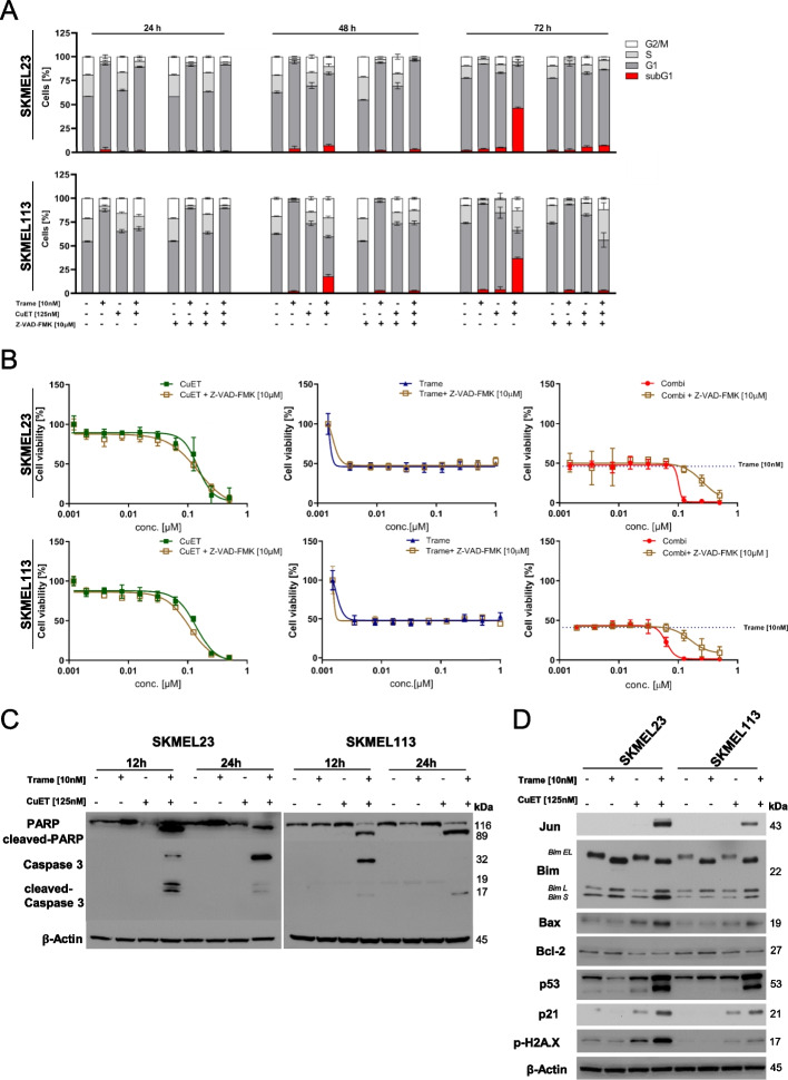
Fig. 3.
Apoptosis induction by trametinib plus CuET in BRAF WT melanoma cells. A BRAF WT melanoma cells were treated with trametinib (10 nM), CuET (125 nM) or their combination in the presence or absence (+/-) of the pan-Caspase inhibitor Z-VAD-FMK (10 µM). Cell cycle analysis at 24, 48 and 72 h was performed, and the cell cycle distribution was plotted with apoptotic cells (sub-G1 fraction) shown as red bars. Three independent experiments were performed (mean ± SD, n = 3 independent experiments). B Cell viability assays (MUH assay) of BRAF WT melanoma cells (SKMEL23 and SKMEL113) after 72 hours of treatment with increasing concentrations of trametinib (up to 2 µM), CuET (up to 500 nM) or their combination with and without (+/-) the pan-Caspase inhibitor Z-VAD-FMK (10 μM). Signals were normalized to untreated controls (n = 3 independent experiments; mean ± SD). Dotted lines represent the effect of 10 nM trametinib on cellular viability. C BRAF WT melanoma cells were treated with trametinib (10 nM), CuET (125 nM) or their combination for 12 and 24 hours. Immunoblots (cropped) of whole cell lysates showing cleavage of Caspase-3 and PARP compared to β-Actin. A representative result of two independent experiments is shown. D Lysates at 12 hours after treatment were prepared, and immunoblot detections for JUN, BIM isoforms (extra-long: EL, long; L, short: S), BAX, BCL-2, p53, p21 and phosphorylated phospho-H2A.X were performed. β-Actin served as a loading control. A representative result of two independent experiments is shown

