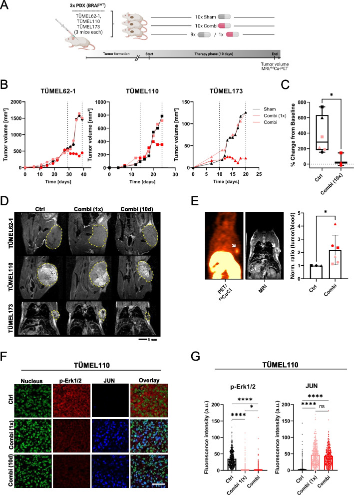
Fig. 7.
The MEK inhibitor trametinib combined with disulfiram impairs BRAF WT melanoma growth in vivo. A Sketch of the NSG mouse experiment with three different patient-derived xenograft (PDX) BRAF WT melanoma models (TÜMEL62-1, TÜMEL110 and TÜMEL173). Three groups were formed: sham treatment; 1x combination treatment (9 days sham and final day combination); and 10x combination treatment (last 10 days combination). DSF (50 mg/kg) plus trametinib (0.3 mg/kg) was orally administered q.d. On the final day, mice received treatment and were subjected to a 64Cu-PET/MR scan. B Tumor growth curves of PDX BRAF WT melanoma models (TÜMEL62-1 [circles], TÜMEL110 [squares] and TÜMEL173 [triangles]) showed diminished tumor growth under combination therapy of trametinib with DSF (red symbols) versus Ctrl mice (sham group, black symbols; 9x sham and 1x combi light red symbols). The dotted lines indicate the therapy phase (Tx) of 10 days. C Statistical analysis (Mann-Whitney test) of the final tumor volumes for the groups: Ctrl (sham mice and 9x sham plus 1x combi mice; n = 6; black and light red symbols); Combi (10x combi; n = 3; red symbols). D T2-weighted magnetic resonance (MR) imaging was performed on the final day. Representative images for visualization of the combination therapy (1x/10x) or sham-treated subcutaneous TÜMEL110 tumors are shown. Tumors are outlined with yellow dashed lines. E Representative PET image of 64CuCl2 uptake in the tumors (shown is TÜMEL173) of combination-treated mice and corresponding MRI image. Normalized tumor-to-blood ratios (sham treated set as 1) showing the specific accumulation of 64CuCl2 in combination-treated PDX tumors (TÜMEL62-1 [circles], TÜMEL110 [squares] and TÜMEL173 [triangles]) in vivo 6 h after radiotracer injection. Tumors show an up to 4-fold increased uptake of the radiotracer compared to the blood (Mann-Whitney test). F Confocal immunofluorescence analysis of phospho-ERK1/2 and JUN in the tumors. Combination therapy for 10 days diminished ERK1/2 phosphorylation and increased JUN protein levels in the tumors of PDX TÜMEL110 (red color: phospho-ERK1/2; blue color: JUN; green: nuclei /Yopro-1; scale bar 50 µm). G Mean fluorescence intensities of phospho-ERK1/2 and JUN staining were used for quantification (≥ 160 cells/group were analyzed). Kruskal-Wallis with subsequent Dunn’s multiple comparisons test. *p< 0.05; ** p < 0.01; *** p < 0.001; and **** p < 0.0001, ns (not significant)

