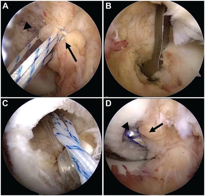
Figure 1.
Intraoperative arthroscopic images demonstrating (A) placement of repair sutures within the residual PCL stump (arrow) avulsed from the femoral origin (arrowhead); (B) drilling of the tibial tunnel with the placement of the drill guide through the medial Gillquist interval; (C) suture configuration during graft passage, with graft, cortical button construct (white sutures), and repair sutures (white/blue stripes) being advanced into the femoral tunnel; and (D) the final construct, with augmentation graft (arrowhead) and native PCL repair (arrow). PCL, posterior cruciate ligament.

