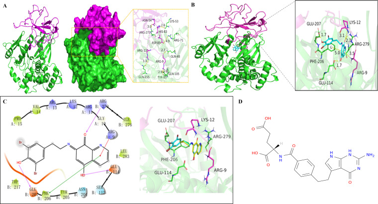
Fig 5.
Screening out the drug Pemetrexed acting on the interaction region of the PEDV N and Ezrin using molecular docking technology. (A) Overall three-dimensional structure and interaction site of PEDV N (purple) and Ezrin (green), with yellow dashed lines representing hydrogen bonds or salt bridges. (B) Overall three-dimensional structure of the PEDV N-Ezrin complex and a close-up view of the active site where the NSC668394 ligand (blue rod) binds to the PEDV N-Ezrin complex. (C) Interaction diagram of NSC668394 ligand (yellow) with the PEDV N-Ezrin complex, with protein residues represented by circles (green, hydrophobic residues; purple, polar residues). (D) The chemical structure of Pemetrexed.

