FIGURE 4.

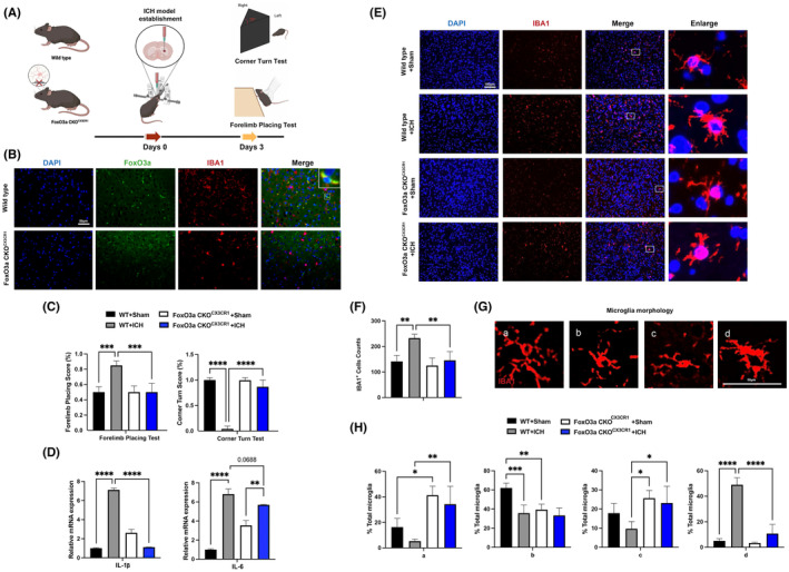
Conditional knockout of FoxO3a in microglia attenuated the neurological deficits and brain injury induced by ICH. (A) Schematic diagram of the experimental design. (B) Representative images showed FoxO3a knockout efficiency in microglia from wild‐type and FoxO3a CKOCX3CR1 mice. (C) The forelimb placing test and corner turn test of the FoxO3afl/fl and FoxO3a cKOCX3CR1 mice in ICH model by injection of autologous whole blood (n = 5). (E) Representative images of IBA1 expression in the striatal area (n = 5). (D) The mRNA levels of IL‐1β and IL‐6 were determined by RT‐PCR in the striatum of the FoxO3afl/fl and FoxO3a cKOCX3CR1 mice caused by ICH (n = 3). (E) Representative pictures of microglia activation phenotype in the striatal region (n = 5). (F) The area of the Iba1+ cells in the striatal region. (G) Morphological microglial analysis was represented with the scale for (H), ranking from a (more ramified phenotype) to d (more amoeboid phenotype) was performed (n = 5). Data were expressed as mean ± SD. *p < 0.05, **p < 0.01, ***p < 0.001.
