
FIGURE 5.
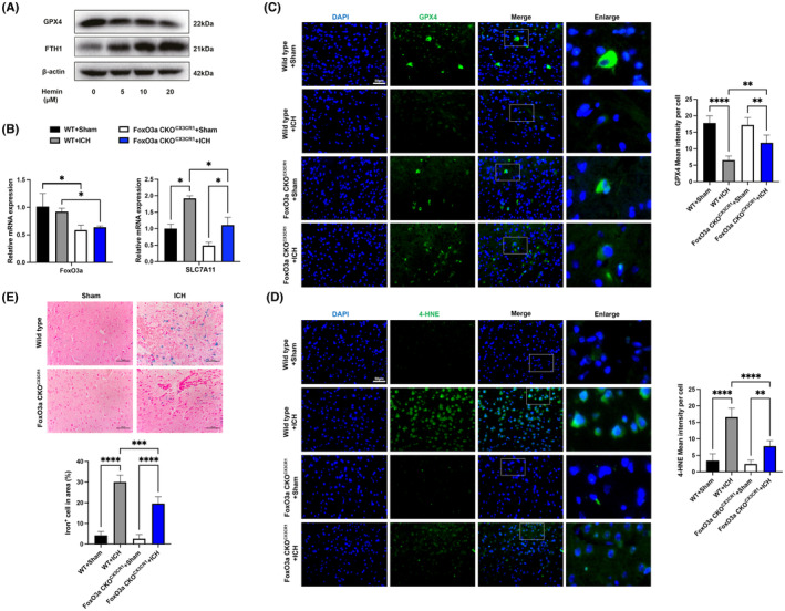
Conditional knockout of FoxO3a in microglia reduces ferroptosis and lipid peroxidation caused by ICH. (A) BV‐2 cells were treated with different concentrations of hemin for 24 h, and then the protein expressions of GPX4 and FHT1 were determined with western blot. (B) The mRNA levels of FoxO3a/SLC7A11 in the striatum of ICH model were determined by qRT‐PCR (n = 3). (C) Representative immunofluorescence images of GPX4 expression (n = 5). (D) Representative immunofluorescence images of 4‐HNE (n = 5) and (E) Representative images of Perl's iron stain in the striatal area of the FoxO3afl/fl and FoxO3a cKOCX3CR1 mice caused by ICH (n = 5). Data were expressed as mean ± SD. *p < 0.05, **p < 0.01, ***p < 0.001.